
The Bronchial Tree: Twenty-Three Generations of Branching
Put your finger on the front of your neck, just below the Adam's apple, and press gently. That firm ridged tube under the skin is the whole of your airway at its beginning — a single pipe about as wide as your thumb. Twenty-three divisions later that one pipe has become roughly 300 million alveoli whose combined surface, spread flat, would cover a tennis court. Nothing designs each of those millions of endings individually. The lung is built by one instruction — split in two — repeated until the branches are thinner than a hair, and every clinical fact worth knowing about breathing falls out of where you are along that sequence.
A two-year-old is sitting on the kitchen floor with a bowl of peanuts while the adults talk. He laughs at something, breathes in at the wrong moment, and a single peanut disappears down his throat. He coughs violently for half a minute — a red-faced, terrifying half minute — and then, alarmingly, stops. He looks fine. He goes back to playing. Three weeks later he is brought to a clinic with a cough that will not clear and a wheeze that only a stethoscope on the right side of his chest can hear, and a chest film shows the right lung looking oddly over-inflated. The peanut is not lost. It is sitting exactly where anatomy said it would sit, in the right main bronchus, because that bronchus is wider, shorter and steeper than its partner, and everything that falls into the airway falls to the right. A bronchoscope retrieves it in twenty minutes. The diagnosis was made by a fact about the angle of a tube.
A tube that must never collapse — and must sometimes be squashed
The trachea solves two opposite problems at once, and its shape is the compromise. The trachea (windpipe) begins at the lower border of the cricoid cartilage, at the level of the C6 vertebra, as the direct continuation of the larynx, and it ends by dividing at the CARINA at the level of the sternal angle — the T4/T5 intervertebral disc. In an adult it is about 10–12 cm long and 2–2.5 cm wide, and it is held permanently open by 15–20 bars of hyaline cartilage. Each bar is not a complete ring but a horseshoe: a C-shaped hoop that is deficient posteriorly. That gap is closed by the TRACHEALIS, a sheet of smooth muscle bridging the free ends of each cartilage, plus fibroelastic tissue. The result is a tube that cannot be crushed shut by the negative pressure of a hard inspiration — which a purely membranous tube certainly would be — and yet has one soft wall. The soft wall faces backwards, and directly behind it, sharing that wall, lies the oesophagus. That single arrangement explains a swallowed bolus bulging forward into the airway on an endoscopic view, the trachealis contracting to narrow the lumen and raise expiratory air speed during a cough, and the uncomfortable anatomical fact that a tracheo-oesophageal fistula — congenital, or eroded through by a tumour or a cuffed tube — has only one thin partition to breach.
The lining is as purposeful as the skeleton. The mucosa is pseudostratified ciliated columnar epithelium — the classic "respiratory epithelium" — studded with goblet cells and supported by submucosal seromucous glands. Together they lay down a two-layer blanket: a watery periciliary layer in which the cilia can beat freely, and a sticky mucus raft floating on top of it that traps dust, bacteria and pollen. Roughly 200 cilia per cell beat about twelve times a second, always upwards, moving that raft towards the larynx at around 1 cm per minute, where it is swallowed without you ever noticing. This is the MUCOCILIARY ESCALATOR, and it is the lung's single most important defence. Blood comes from the inferior thyroid arteries above and bronchial branches of the thoracic aorta below; venous blood drains to the inferior thyroid veins; lymph goes to the pretracheal and paratracheal nodes. Nerve supply is from the vagus and its recurrent laryngeal branches — sensory, secretomotor and bronchoconstrictor — with sympathetic fibres from the sympathetic trunk, all discussed in the nerves of the thorax.
The carina, and the asymmetry that decides where things land
The last cartilage of the trachea is also the most sensitive square centimetre of the airway. At the level of the sternal angle — the same landmark that marks rib 2, the T4/T5 disc, the beginning and end of the aortic arch and the division of superior from inferior mediastinum, as set out in the thoracic cage — the last tracheal cartilage becomes a sharp sagittal ridge projecting upwards between the two orifices below it. This is the carina, and to a bronchoscopist it is the single most important landmark in the chest: a crisp, knife-edge carina means normal anatomy, while a broadened, splayed carina means something is pushing it apart from below, classically enlarged subcarinal lymph nodes in bronchial carcinoma. It is also the most sensitive cough-reflex point in the whole airway; touching it with a suction catheter in a conscious patient provokes an immediate, violent cough, mediated by the vagus. Note the deep breath, glottic closure, rise in intrathoracic pressure and explosive release that follow — and note that the trachealis narrows the tube at that instant, so the same volume of air leaves through a smaller aperture and reaches a velocity approaching that of a hurricane.
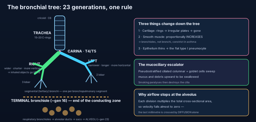
Below the carina the two main (primary) bronchi are emphatically not mirror images. The RIGHT main bronchus is WIDER, SHORTER (about 2.5 cm before it gives its first branch) and MORE VERTICAL, leaving the trachea at roughly 25 degrees from the vertical. The LEFT is narrower, longer (about 5 cm) and far more horizontal, at roughly 45 degrees, because it must travel across the midline beneath the arch of the aorta and in front of the oesophagus to reach a lung whose space is partly taken by the heart. The consequence is one of the most reliably examined facts in medicine, and it is not merely academic: an inhaled peanut or tooth, aspirated vomit or gastric contents, and an endotracheal tube advanced a centimetre or two too far ALL go right. Advance the tube far enough and it intubates the right main bronchus alone, ventilating one lung while the left collapses — which is why the tube's depth is checked, the chest is auscultated bilaterally, and the film is reviewed. Gravity then decides the segment: in an upright patient material tends to the posterior basal segment of the right LOWER lobe; in a patient lying supine — the anaesthetised, the obtunded, the drunk — it settles in the posterior segment of the right UPPER lobe, which is exactly where the aspiration abscess of an unconscious patient appears on the film.
Think of the airway as a motorway system draining a country. The trachea is the single national motorway leaving the capital: wide, rigid, engineered never to close. It splits into two trunk roads, then into three lanes on the right and two on the left, then into ten regional routes per side, and on down through county roads, town streets and finally the cul-de-sacs where people actually live. Two things happen as you descend. Each individual road gets narrower — but there are so many more of them that the TOTAL width of tarmac at every level grows enormously, so traffic that roared out of the capital is barely crawling by the time it reaches the driveways. And the roads change construction: the motorway is reinforced concrete (cartilage), the town streets have no reinforcement at all and are lined instead with muscular walls that can squeeze the street narrower (the bronchioles). Close the motorway and everything stops at once. Squeeze ten thousand small streets by a third each, and the capital never notices until the whole country is gridlocked — which is precisely what an asthma attack is.
The branching order, named precisely
Every name in the chain tells you what territory that branch owns. The sequence, in order: main (primary) bronchi → LOBAR (secondary) bronchi, one to each lobe, so THREE on the right and TWO on the left → SEGMENTAL (tertiary) bronchi, each supplying one bronchopulmonary segment → smaller conducting bronchioles → TERMINAL bronchioles, the last purely conducting airway, at about generation 16 → RESPIRATORY bronchioles, whose walls begin to carry occasional alveoli → alveolar ducts → alveolar sacs → alveoli. Everything from the nose to the terminal bronchiole is the CONDUCTING zone: it moves air and conditions it, but no gas exchange occurs there — this is the anatomical dead space, about 150 mL in an adult, which is why shallow, rapid panting ventilates the dead space and not the lung. Everything from the respiratory bronchiole onwards is the RESPIRATORY zone, where blood and air are separated only by the fused basement membranes of the alveolar epithelium and the capillary endothelium, some 0.2–0.6 micrometres of tissue, as traced in pulmonary circulation and breathing mechanics.
The segmental bronchi deserve special attention, because each one, together with its accompanying branch of the pulmonary artery, defines a BRONCHOPULMONARY SEGMENT: a pyramidal wedge of lung with its apex at the hilum, its own air supply, its own arterial supply, and — crucially — connective tissue septa separating it from its neighbours, with the pulmonary veins running in those septa between segments rather than within them. There are ten on the right and, because of fusion, usually eight to ten on the left. This is not trivia. It is the reason a surgeon can remove a single diseased segment and leave the rest of the lobe intact, the reason a localised abscess or a tuberculous focus tends to stay within one segment's borders on a CT, and the reason physiotherapists position patients so that a given segment's bronchus points downhill for postural drainage. The lobes and fissures those segments assemble into are the subject of the lungs.
What changes as you descend the tree
Four gradients run down the airway at once, and every one of them has a clinical consequence. First, cartilage. The neat C-rings of the trachea become irregular, interlocking plates in the bronchi — plates that are needed because these tubes are no longer a simple cylinder — and then, at a diameter of about 1 mm, cartilage disappears entirely. By definition, an airway with no cartilage in its wall is a BRONCHIOLE. Second, smooth muscle. As cartilage falls away, a complete spiral coat of smooth muscle takes over the job of holding and controlling the calibre, so muscle makes up a proportionally greater share of the wall the further down you go. Third, epithelium. Pseudostratified ciliated columnar with goblet cells gives way to simple columnar, then to simple cuboidal in the terminal bronchioles, and finally to the extraordinary flattened type I pneumocyte of the alveolus, so thin that gas crosses it almost as though it were not there. Fourth, glands and cell types. Submucosal glands and goblet cells vanish below the bronchioles — mucus in the alveoli would be a catastrophe — and in their place appear club (formerly Clara) cells, which secrete a watery surfactant-like protein, detoxify inhaled substances and act as progenitor cells for repair; while in the alveolar wall the cuboidal type II pneumocyte manufactures true surfactant and serves as the stem cell for type I.
Alongside those structural gradients runs a purely geometrical one that governs the physics of breathing. Every division roughly doubles the number of tubes while reducing each one's diameter by much less than half, so the TOTAL cross-sectional area of the airway increases with staggering speed — from about 2.5 square centimetres at the trachea to several thousand square centimetres by the alveolar ducts. Since flow rate is fixed but area explodes, air velocity collapses: what leaves the trachea at metres per second is barely moving at all in the terminal bronchioles, and the last part of the journey into the alveolus is accomplished not by bulk flow but by diffusion, which is fast enough only because the distance has become tiny. There is a second surprise hidden in the same arithmetic. Because resistance falls as the total area rises, most of the resistance of the normal airway lies not in the small tubes but in the medium-sized bronchi, roughly the first eight generations. The small airways are a "silent zone" — they can be substantially diseased before ordinary spirometry notices.
Ask where an asthma attack physically happens and the answer is not "the bronchi" — it is the BRONCHIOLES, and the reason is architectural. A bronchus has cartilage plates in its wall; no matter how hard the smooth muscle contracts, the cartilage will not let it close. A bronchiole has no cartilage at all and a proportionally thicker muscular coat, so its calibre is set entirely by muscle tone, and it can be narrowed dramatically, or shut. Add mucosal oedema and mucus plugging and the airway that was already the smallest closes first. That is why the wheeze of asthma is EXPIRATORY — during expiration the surrounding lung pressure rises and squeezes the unsupported bronchioles further shut, whereas inspiration pulls them open. It is also why bronchodilators work: they relax exactly the muscle that has no cartilage arguing with it, in asthma and COPD alike.
The escalator, and everything that breaks it
The mucociliary escalator runs day and night and you never feel it. Break it and the lung's defence collapses in a predictable order. Cigarette smoke first paralyses the cilia and then destroys them, while simultaneously driving goblet-cell hyperplasia — more mucus, and nothing to move it. That is the mechanism of the smoker's morning cough: mucus accumulated overnight while the escalator was off, cleared by brute force on waking. In bronchiectasis the cycle closes on itself: infection damages the wall, the damaged wall fails to clear secretions, retained secretions breed infection, and the bronchi are permanently, irreversibly dilated, producing cupfuls of purulent sputum. In cystic fibrosis the periciliary fluid layer is dehydrated so the mucus raft is too thick to be moved at all; in primary ciliary dyskinesia the cilia are structurally abnormal and simply do not beat — the same defect that leaves sperm immotile and, in Kartagener's syndrome, allows the viscera to end up mirror-reversed. Chronic irritation can also drive squamous metaplasia, ciliated epithelium replaced by flat protective squamous cells that clear nothing, a change that is reversible on stopping smoking but is also the first step on the road to carcinoma.
The tube that went too far: a patient is intubated in the emergency department and the chest rises well, but only on the right; auscultation is silent on the left and the film shows a collapsed left lung. The tube has slipped past the carina into the right main bronchus. Withdraw it two centimetres and both lungs inflate. The hole in the neck: an emergency cricothyroidotomy is made through the cricothyroid membrane because it is subcutaneous and bloodless, but a planned tracheostomy is placed lower, through the second to fourth tracheal rings, avoiding the first ring (to protect the cricoid from stenosis) and staying above the thyroid isthmus and the great veins. The inhaler that beats the tablet: a salbutamol puff reaches the bronchiolar smooth muscle in seconds at a fraction of a milligram, whereas a tablet must be absorbed, circulate and bathe the whole body — the anatomical argument for delivering airway drugs by inhalation. The trachea that moved: a young man with a tension pneumothorax arrives breathless, and the trachea, palpable in the suprasternal notch, is pushed away from the affected side — whereas in a collapsed, fibrosed lobe it is pulled towards it. One finger in the notch, two opposite diagnoses.
- The trachea runs from the lower border of the cricoid cartilage (C6) to the carina at the sternal angle (T4/T5); 10–12 cm long, 2–2.5 cm wide, held open by 15–20 C-shaped hyaline cartilage rings.
- The rings are deficient POSTERIORLY, where the trachealis smooth muscle bridges them and the oesophagus lies directly behind — hence a bolus bulging forward, cough-time narrowing, and the ease of a tracheo-oesophageal fistula.
- The lining is pseudostratified ciliated columnar epithelium with goblet cells: the mucociliary escalator, sweeping mucus upwards to the larynx — paralysed and then destroyed by cigarette smoke.
- Blood from the inferior thyroid arteries (and bronchial arteries below); nerves from the vagus and recurrent laryngeal nerves plus sympathetic fibres; lymph to pretracheal and paratracheal nodes.
- The carina is the last tracheal cartilage, the most sensitive cough-reflex point, and the key bronchoscopic landmark — a splayed carina suggests enlarged subcarinal nodes.
- The RIGHT main bronchus is wider, shorter (~2.5 cm) and more vertical (~25°) than the left (~5 cm, ~45°) — so foreign bodies, aspirate and an over-inserted endotracheal tube all go RIGHT.
- Branching order: main (primary) → lobar (secondary; 3 right, 2 left) → segmental (tertiary) → conducting bronchioles → terminal bronchioles → respiratory bronchioles → alveolar ducts → alveolar sacs → alveoli.
- Everything down to the TERMINAL bronchiole (generation ~16) is the conducting zone — the anatomical dead space, ~150 mL. Gas exchange begins at the RESPIRATORY bronchiole.
- Each segmental bronchus plus its pulmonary artery branch defines a bronchopulmonary segment (10 right, 8–10 left), separated by septa carrying the pulmonary veins — the basis of segmentectomy and postural drainage.
- Down the tree: cartilage rings → irregular plates → none (that absence DEFINES a bronchiole); smooth muscle proportionally increases; epithelium thins from pseudostratified to cuboidal to the type I pneumocyte.
- Glands and goblet cells disappear distally; club (Clara) cells appear in the bronchioles, and type II pneumocytes in the alveoli make surfactant and regenerate type I cells.
- Total cross-sectional area grows enormously with each generation, so air velocity collapses and the last step into the alveolus is by diffusion; most normal airway resistance is in medium bronchi, leaving the small airways a "silent zone".
- Saying the tracheal cartilages are complete rings. They are C-shaped and deficient posteriorly — the trachealis muscle completes them. A truly complete ring would leave no room for the oesophagus to expand behind it.
- Placing the tracheal bifurcation at the level of the manubrium or the neck. The carina lies at the STERNAL ANGLE, the T4/T5 disc — the same plane as rib 2 and the limits of the aortic arch.
- Blaming an asthma attack on the bronchi. Bronchi have cartilage that resists closure; it is the cartilage-free, muscle-rich BRONCHIOLES that constrict — which is also why the wheeze is expiratory.
An unconscious patient is intubated in the emergency department. The chest wall rises on the right only, breath sounds are absent on the left, and the film shows a collapsed left lung with the endotracheal tube tip well below the level of the sternal angle. What is the anatomical explanation?
- The trachea runs from the cricoid cartilage (C6) to the carina at the sternal angle (T4/T5), 10–12 cm of tube held open by 15–20 C-shaped cartilages that are deficient posteriorly, where the trachealis muscle and the oesophagus lie.
- Its pseudostratified ciliated epithelium and goblet cells form the mucociliary escalator, the lung's chief defence — paralysed by smoking, defeated in cystic fibrosis and primary ciliary dyskinesia, and trapped in the vicious circle of bronchiectasis.
- The right main bronchus is wider, shorter and more vertical, so inhaled objects, aspirated material and over-inserted tubes go right — to the right lower lobe when upright, the posterior segment of the right upper lobe when supine.
- Main → lobar → segmental → bronchioles → terminal bronchioles (the end of the conducting zone) → respiratory bronchioles → alveolar ducts → sacs → alveoli; along the way cartilage vanishes, smooth muscle takes over (so bronchioles, not bronchi, constrict in asthma), the epithelium thins to the type I pneumocyte, and the total cross-sectional area grows until air arrives by diffusion alone.
- Drake RL, Vogl AW, Mitchell AWM. Gray's Anatomy for Students — Thorax: trachea, bronchi and the bronchial tree.
- Moore KL, Dalley AF, Agur AMR. Clinically Oriented Anatomy — The trachea, main bronchi and bronchopulmonary segments.
- Netter FH. Atlas of Human Anatomy — Trachea and major bronchi; intrapulmonary airways.
- Last RJ. Last's Anatomy: Regional and Applied — Trachea and lungs.
- Snell RS. Clinical Anatomy by Regions — Foreign body inhalation, tracheostomy and bronchoscopic anatomy.
- TeachMeAnatomy — The Trachea; The Bronchi; Bronchopulmonary Segments.

