
Nerves of the Thorax: Long Journeys and Strange Detours
Most nerves take the shortest route to what they supply. The thorax contains two that do not. One is born in the neck, at the level of the third, fourth and fifth cervical segments, and then travels the whole length of the chest to reach the muscle that makes you breathe — because that muscle began life in the neck and dragged its nerve downwards as the embryo grew. The other dives into the chest, hooks under a great artery, turns around and climbs all the way back up to the throat, adding a detour of many centimetres for no reason a designer would accept. Neither route makes engineering sense. Both make perfect sense as history — and both explain a set of clinical signs that are impossible to reason out any other way.
A fifty-eight-year-old schoolteacher comes to the clinic because his voice has gone. Not painfully — there is no sore throat, no fever, nothing that behaves like an infection. It simply turned husky about three months ago and never came back, and by the last period of the day he cannot make himself heard across the classroom. He has smoked since he was seventeen. The doctor listens to the story, looks at the man's neck, finds nothing at all there, and then does something that seems to have nothing to do with a voice: she orders a chest X-ray. It shows a mass at the left hilum. The tumour never touched the larynx. It grew around a nerve passing under the arch of the aorta, five inches below the vocal cords, on its way back up to the throat — a nerve whose route through the mediastinum was set five weeks after conception and has cost people their voices ever since.
One landmark sorts them all: the root of the lung
If you remember only one sentence about thoracic nerves, remember this one. The phrenic nerve passes ANTERIOR to the root of the lung; the vagus nerve passes POSTERIOR to it. That single relationship is the discriminator that surgeons, radiologists and examiners all use, and it is not an arbitrary fact to memorise — it follows from where each nerve is going. The phrenic is heading for the diaphragm along the side of the pericardium, and the pericardium lies in front of the hila, so the phrenic must run in front. The vagus is heading for the oesophagus and the posterior mediastinum, both of which lie behind the hila, so the vagus must run behind. Everything else in this article hangs off that geometry: which structures each nerve is plastered against, which tumours reach it first, and which operations put it at risk.
The phrenic nerve: C3, 4 and 5 keep the diaphragm alive
"C3, 4, 5 keeps the diaphragm alive" is the most useful rhyme in anatomy — and a spinal-injury prognosis in six words. The phrenic nerve arises from the cervical plexus, chiefly from the anterior ramus of C4 with contributions from C3 and C5. It forms at the upper lateral border of the anterior scalene muscle and descends almost vertically across its front, running from lateral to medial under the prevertebral fascia, and enters the thorax by passing between the subclavian artery and the subclavian vein. From there the two nerves separate in character. The right phrenic is short and straight: it descends on the right brachiocephalic vein, then the superior vena cava, then the right side of the pericardium over the right atrium. The left phrenic is longer: it crosses the left side of the aortic arch — superficial to the left vagus, an important relation — passes in front of the left lung root and runs down over the bulge of the left ventricle. Both nerves travel in the same tunnel: between the fibrous pericardium and the mediastinal pleura, accompanied throughout by the pericardiacophrenic artery and vein.
The phrenic is a mixed nerve doing two very different jobs. It is the SOLE motor supply to its own half of the diaphragm — cut it and that hemidiaphragm is paralysed, whatever the intercostal muscles do. And it is sensory to a surprisingly wide territory: the fibrous pericardium and the parietal serous pericardium, the mediastinal pleura, the diaphragmatic pleura and the central diaphragmatic peritoneum on its own side. That sensory list is why irritation anywhere along it — blood under the diaphragm, a subphrenic abscess, gallbladder inflammation touching the diaphragmatic peritoneum, pericarditis — produces pain not in the chest but at the TIP OF THE SHOULDER, referred to the C3–C4 dermatome served by the supraclavicular nerves. The two nerves also end differently: the right phrenic reaches the diaphragm and pierces it at the caval opening at the T8 level, travelling with the inferior vena cava, while the left pierces the muscular part of the diaphragm separately, just left of the pericardium.
The vagus nerves: the great parasympathetic highway
The tenth cranial nerve is the only one that leaves the head and keeps going until it runs out of gut. Each vagus (CN X) enters the thorax inside the carotid sheath and immediately reveals a right–left difference. The right vagus descends on the right side of the trachea, behind the right brachiocephalic vein and the superior vena cava, and passes BEHIND the root of the right lung. The left vagus enters between the left common carotid and left subclavian arteries, crosses the LEFT SIDE OF THE AORTIC ARCH (with the left phrenic superficial to it), and passes BEHIND the root of the left lung. On the way each contributes fibres to three mixed autonomic networks — the cardiac plexus, the pulmonary plexus and the oesophageal plexus — then breaks up into a meshwork on the oesophagus and re-gathers below it. Because the stomach rotates during development, dragging its nerves with it, the LEFT vagus ends up as the ANTERIOR vagal trunk and the RIGHT as the POSTERIOR trunk. Both pass with the oesophagus through the T10 hiatus into the abdomen. The mnemonic students never forget is LARP: Left Anterior, Right Posterior.
Functionally the vagus is the parasympathetic supply to everything in the chest and to the gut as far as the distal third of the transverse colon. In the heart it slows the sinoatrial node and delays conduction through the atrioventricular node — the resting brake that keeps a healthy pulse near seventy rather than the intrinsic pacemaker rate of about a hundred. In the airways it constricts bronchial smooth muscle and increases glandular secretion, which is exactly why muscarinic antagonists are useful inhaled bronchodilators. In the oesophagus and stomach it drives peristalsis and secretion. It also carries a large afferent load upwards: stretch information from the lungs, chemoreceptor and baroreceptor traffic, and the visceral sensation that makes a distended oesophagus feel like nothing you can localise.
The recurrent laryngeal nerves: the detour that embryology forced
Two nerves with the same job, the same origin and wildly different lengths. In the embryo each recurrent laryngeal nerve is the nerve of the sixth pharyngeal arch, and it lies caudal to the sixth arch artery on its own side. Then the neck elongates and the heart descends. On the RIGHT, the distal sixth arch artery regresses entirely and the fifth disappears too, so the nerve is left hooked around the next artery still standing above it — the fourth arch derivative, the RIGHT SUBCLAVIAN ARTERY — and it stays in the neck. On the LEFT, the sixth arch artery persists as the ductus arteriosus, which after birth becomes the LIGAMENTUM ARTERIOSUM, and this tethers the left nerve to the underside of the arch of the aorta. So the left nerve is dragged deep into the thorax, hooks under the arch just lateral to the ligamentum arteriosum, and then climbs back up in the groove between the trachea and the oesophagus to reach the larynx. It is several times longer than its partner. Both nerves supply ALL the intrinsic muscles of the larynx except cricothyroid (which belongs to the external branch of the superior laryngeal nerve) and carry sensation from below the vocal folds.
That extra length is not a curiosity; it is a list of diseases. Because the left recurrent laryngeal nerve spends its journey inside the mediastinum, it can be compressed or infiltrated by anything that grows there: a left hilar bronchial carcinoma, an aneurysm of the aortic arch, enlarged mediastinal lymph nodes, and — classically — a hugely dilated left atrium in mitral stenosis pressing upwards on the nerve, a combination called Ortner's or cardiovocal syndrome. Any of these produces the same sign, HOARSENESS, from paralysis of the vocal cord on that side. The right nerve, hooking only around the right subclavian artery high in the neck, is out of the mediastinum's reach; it is instead at risk from thyroid surgery, and from the rare "non-recurrent" right nerve that runs straight in from the vagus when an aberrant right subclavian artery has changed the embryology. Hoarseness that appears without a sore throat and lasts more than three weeks is therefore a red flag, not a voice complaint.
Imagine a cable running from an office on the ground floor of a building to a socket a metre away, looped for tidiness under a steel beam. Now the building is rebuilt around it: the beam is lowered five storeys and the cable, still hooked underneath, is dragged down with it — and to reach the same socket a metre from where it started it must now run down five floors, round the beam, and back up again. Nobody would install it that way. But nobody installed it; it was inherited from a smaller building. The left recurrent laryngeal nerve is that cable, the beam is the sixth aortic arch turned ligamentum arteriosum, and the descent of the heart is the rebuilding. In the giraffe, the same detour is over four metres long.
A voice is a chest sign. Persistent hoarseness with no throat pathology means: examine the vocal cords, and if one is paralysed, image the mediastinum — because the nerve that moves it has already been down there. In practice a new, painless, progressive hoarseness in a smoker is one of the classic presentations of bronchial carcinoma, and the tumour that causes it is often on the left, near the hilum, nowhere near the larynx. The lesson generalises: a nerve's clinical territory is not where it ends, it is everywhere it has travelled.
The sympathetic trunk and the splanchnic nerves
A beaded chain lying on the rib heads, just under the pleura, running the whole length of the chest. The sympathetic trunk is a paired chain of ganglia linked by nerve fibres, running down each side of the vertebral column. In the upper thorax it lies over the HEADS OF THE RIBS; lower down, as the ribs slope, it moves onto the sides of the vertebral bodies, and it is covered only by parietal pleura — which is why it is visible at thoracoscopy and reachable with a diathermy hook. There are usually eleven or twelve thoracic ganglia, and the first is commonly fused with the inferior cervical ganglion to form the cervicothoracic (stellate) ganglion at the neck of the first rib. Each ganglion exchanges two kinds of connection with its spinal nerve: WHITE rami communicantes carry myelinated preganglionic fibres into the chain (these exist only from T1 to L2, the thoracolumbar outflow of the segmental nerves), while GREY rami carry unmyelinated postganglionic fibres out to every spinal nerve, supplying sweat glands, arrector pili and blood vessels throughout the body wall.
Some preganglionic fibres refuse to synapse in the chain at all and pass straight through it, gathering into the SPLANCHNIC nerves that dive into the abdomen. The GREATER splanchnic nerve comes from ganglia T5 to T9 and synapses in the coeliac ganglion. The LESSER splanchnic nerve comes from T10 and T11 and synapses in the aorticorenal ganglion. The LEAST splanchnic nerve, when present, comes from T12 and joins the renal plexus. All three pierce the crura of the diaphragm to reach their targets — which is why the sympathetic supply of the foregut and midgut is set up entirely inside the chest. Meanwhile the cardiac sympathetic supply arises higher, from segments T1 to T4 or T5, reaching the heart through cervical and upper thoracic cardiac branches; these fibres accelerate the heart, increase the force of contraction and dilate the coronary arteries — the counterweight to the vagus, and the whole fight-or-flight versus rest-and-digest balance in one organ.
That segmental arrangement explains the most important referred pain in medicine. Visceral afferent fibres from the heart travel back with the sympathetic nerves and enter the spinal cord at T1 to T4/T5 — the very same segments that receive somatic sensation from the chest wall and the medial side of the arm. The brain, which has never in a lifetime received a message from the myocardium and has no map for it, misreads the traffic as coming from the body wall it knows. So myocardial ischaemia is felt as crushing central chest pain radiating to the left arm, and often to the neck and jaw, where convergence of vagal afferents onto the same central neurons carries the signal higher still. Nothing is wrong with the arm; the wiring simply arrives at a shared address. The pulmonary, cardiac and oesophageal plexuses that lie around the tracheal bifurcation and the great vessels are the mixing points where all of this comes together — sympathetic fibres from the chain and parasympathetic fibres from the vagus, woven into single networks that no dissection can cleanly separate.
The hiccup that would not stop: a man with a subphrenic abscess after appendicitis hiccups for four days and complains of right shoulder-tip pain — one nerve, the phrenic, reporting both the spasm and the pain. The raised diaphragm: a woman is breathless three weeks after coronary artery bypass grafting; her chest X-ray shows a high left hemidiaphragm that moves paradoxically on sniffing — phrenic injury from cold cardioplegia, a recognised complication of cardiac surgery. The drooping eyelid: a smoker with shoulder pain has a small pupil, a partially closed eyelid and a dry face on the same side — Horner's syndrome from a Pancoast tumour at the lung apex eroding into the sympathetic chain and the stellate ganglion. The racing heart: a young man with a regular narrow-complex tachycardia at 190 is asked to blow hard against a closed glottis; vagal tone rises, the atrioventricular node blocks, and the rhythm breaks — the vagus used as a drug, before adenosine is even drawn up. The sweating hands: a student whose palms drip so badly that paper sticks to them has a thoracoscopic sympathectomy, the chain divided over the rib heads at T2–T3; the hands go dry the same day, at the price of compensatory sweating elsewhere.
- The phrenic nerve passes ANTERIOR to the root of the lung; the vagus passes POSTERIOR to it. This one relationship separates them everywhere in the chest.
- Phrenic = C3, C4, C5 (mainly C4), from the cervical plexus, down the front of anterior scalene, between the subclavian artery and vein, then between the fibrous pericardium and mediastinal pleura with the pericardiacophrenic vessels.
- Phrenic is the SOLE motor nerve of its own hemidiaphragm, and sensory to fibrous pericardium, mediastinal and diaphragmatic pleura and central diaphragmatic peritoneum — hence referred pain at the shoulder tip (C3–C4).
- The right phrenic pierces the diaphragm at the caval opening (T8) with the IVC; the left pierces the muscle separately, just left of the pericardium.
- The right vagus passes behind the right lung root; the left crosses the left side of the aortic arch (deep to the left phrenic) and passes behind the left lung root.
- After oesophageal rotation: LEFT vagus → ANTERIOR trunk, RIGHT vagus → POSTERIOR trunk (LARP); both pass through the T10 hiatus with the oesophagus.
- Vagal (parasympathetic) effects in the chest: slows the SA node and AV conduction, constricts bronchi, increases glandular secretion, drives oesophageal peristalsis.
- LEFT recurrent laryngeal hooks under the ARCH OF THE AORTA at the ligamentum arteriosum and climbs the tracheo-oesophageal groove; the RIGHT hooks under the right subclavian artery in the neck.
- Both recurrent laryngeal nerves supply ALL intrinsic laryngeal muscles EXCEPT cricothyroid; the left is vulnerable to left hilar carcinoma, aortic arch aneurysm, mediastinal nodes and a dilated left atrium (Ortner's) → hoarseness.
- The sympathetic trunk lies over the rib heads under the parietal pleura, gives WHITE rami (T1–L2, preganglionic in) and GREY rami (postganglionic out to every spinal nerve).
- Splanchnics pierce the crura: GREATER T5–T9 → coeliac ganglion; LESSER T10–T11 → aorticorenal ganglion; LEAST T12 → renal plexus.
- Cardiac sympathetic outflow is T1–T4/T5, sharing spinal segments with the chest wall and medial arm — the anatomical basis of referred cardiac pain to chest, left arm and jaw.
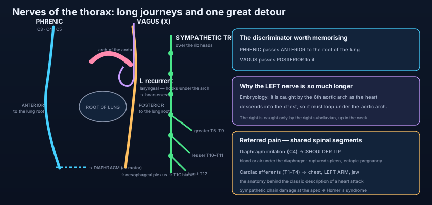
- Swapping the lung-root relations. The PHRENIC is anterior and the VAGUS posterior — the reverse is the single commonest error in thoracic anatomy, and it inverts every clinical prediction that follows from it.
- Thinking hoarseness means a laryngeal problem. The paralysed cord may be innocent; the lesion is often centimetres lower, in the mediastinum, on the left recurrent laryngeal nerve.
- Assuming the recurrent laryngeal nerves supply every laryngeal muscle. They supply all the intrinsic muscles EXCEPT cricothyroid, which belongs to the external branch of the superior laryngeal nerve — which is why the two nerves give different voice changes.
A 60-year-old smoker has had a painless, progressively hoarse voice for two months. Laryngoscopy shows a paralysed LEFT vocal cord. Where is the lesion most likely to be, and why on the left?
- The discriminator: the PHRENIC nerve (C3, C4, C5 — mainly C4) runs ANTERIOR to the root of the lung between the fibrous pericardium and mediastinal pleura with the pericardiacophrenic vessels; the VAGUS runs POSTERIOR to it.
- The phrenic is the sole motor supply to its hemidiaphragm and sensory to fibrous pericardium, mediastinal and diaphragmatic pleura and central peritoneum — hence shoulder-tip referred pain, hiccups from irritation, and paralysis after cardiac surgery; the right pierces the caval opening at T8, the left the muscle separately.
- The vagi form the cardiac, pulmonary and oesophageal plexuses, slow the heart, constrict bronchi and increase secretion; after oesophageal rotation the LEFT becomes the ANTERIOR trunk and the RIGHT the POSTERIOR trunk, both passing the T10 hiatus.
- The LEFT recurrent laryngeal nerve hooks under the aortic arch at the ligamentum arteriosum (an embryological legacy of the sixth aortic arch) and is therefore long and mediastinal — hoarseness in left hilar cancer, aortic aneurysm and mitral stenosis — while the sympathetic trunk lies on the rib heads giving grey and white rami and the greater (T5–T9), lesser (T10–T11) and least (T12) splanchnic nerves, with cardiac outflow from T1–T4/T5 explaining referred cardiac pain.
- Drake RL, Vogl AW, Mitchell AWM. Gray's Anatomy for Students — Thorax: nerves of the mediastinum.
- Moore KL, Dalley AF, Agur AMR. Clinically Oriented Anatomy — The thorax: phrenic and vagus nerves, sympathetic trunks and splanchnic nerves.
- Netter FH. Atlas of Human Anatomy — Nerves of the thorax; autonomic nerves of the thoracic viscera.
- Last RJ. Last's Anatomy: Regional and Applied — The mediastinum and its nerves.
- Snell RS. Clinical Anatomy by Regions — Recurrent laryngeal nerve palsy and referred cardiac pain.
- TeachMeAnatomy — The Phrenic Nerve; The Vagus Nerve; The Sympathetic Trunk.

