
The Pudendal Nerve: S2, S3, S4 — Keeping the Pelvis Off the Floor
One nerve carries the voluntary control of both sphincters, the sensation of the entire perineum, and the reflexes on which continence depends. Sever it and a person loses control of bladder and bowel, loses feeling from the skin between the thighs, and loses the reflex arcs a neurologist uses to test whether a damaged spinal cord is still connected at its lowest levels. And this nerve, carrying all of that, takes one of the strangest routes in the human body: it leaves the pelvis altogether through one hole, hooks around a ligament in the open, and comes straight back in through another hole a centimetre away. That pointless-looking detour is not an accident. It is the reason a midwife can anaesthetise a perineum with a finger and a needle, and the reason a cyclist's saddle can make a limb of the body go numb.
It is two in the morning on a delivery unit. The second stage has gone on too long, the head is on the perineum and will not advance, and the decision has been made to help it out with forceps. There is no time for a regional block to be sited from scratch. The obstetrician does something that looks almost primitive: she slides two fingers into the vagina and walks them backwards and laterally along the wall until she meets a hard, sharp point of bone — the ischial spine. She keeps one fingertip on it. With the other hand she passes a long needle along her fingers and injects a few millilitres of local anaesthetic just medial to that point of bone, then repeats it on the other side. Within ten minutes the perineum, the vulva and the anal margin are numb, the forceps go on without protest, and the episiotomy and its repair are painless. Nothing was scanned and nothing was measured. She found one bony landmark with a fingertip, and through it a nerve she could not see — because that nerve is obliged, by its own peculiar course, to pass exactly there.
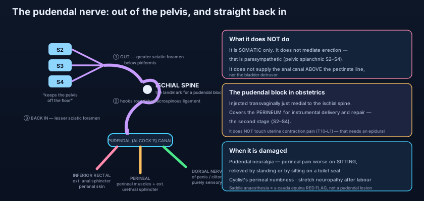
S2, 3, 4 keeps the pelvis off the floor
The mnemonic is crude, but it is the single most useful line in pelvic neuroanatomy. The pudendal nerve arises from the ANTERIOR RAMI of the second, third and fourth sacral nerves — S2, S3 and S4 — as they emerge from the anterior sacral foramina and converge on the anterior surface of the piriformis muscle. There, on that broad muscular sheet, the roots gather into the SACRAL PLEXUS, described in full in the lumbosacral plexus, and the pudendal nerve is one of its named branches, usually formed from the lower divisions of the plexus and lying medial and inferior to the far bulkier sciatic nerve. It is the principal SOMATIC nerve of the perineum: motor to striated muscle, sensory to skin, and carrying the afferent limbs of the pelvic reflexes. "S2, 3, 4 keeps the pelvis off the floor" is worth learning verbatim, because those same three segments also supply the levator ani through the nerve to levator ani, and the same three segments carry the parasympathetic outflow of the pelvis. Injure the cord or the cauda equina at that level and the floor of the pelvis — muscular, sensory and reflex — fails together.
A course that leaves the pelvis only to come straight back in
Follow it in three stages and the whole of its clinical behaviour falls out of the geometry. STAGE ONE — it leaves. The nerve passes out of the pelvis through the GREATER SCIATIC FORAMEN, running BELOW the piriformis muscle, in company with the INTERNAL PUDENDAL ARTERY, which will accompany it for the rest of its journey and give branches that mirror its own. For a few centimetres the nerve is in the gluteal region, outside the pelvis altogether. STAGE TWO — it turns. It crosses the SACROSPINOUS LIGAMENT immediately MEDIAL to the ISCHIAL SPINE, the nerve lying against the ligament with the artery lateral to it. This is the pivot of the whole course, and it matters for one blunt reason: the ischial spine is palpable — through the vagina, or through the rectum — and it is the only landmark in the region that can be found reliably with a fingertip. A needle placed just medial to it is placed on the nerve. STAGE THREE — it re-enters. Having hooked round the ligament, it passes back into the perineum through the LESSER SCIATIC FORAMEN and enters the PUDENDAL CANAL, also called ALCOCK'S CANAL: a fascial sheath in the obturator internus fascia on the LATERAL WALL of the ischioanal fossa, carrying the nerve and the internal pudendal vessels forwards towards the perineal pouches. It is a tunnel with unyielding walls, and that fact alone generates a whole pain syndrome.
Picture a service pipe that has to reach a basement room, but the wall between the corridor and that room is solid concrete with no opening anywhere along it. The only way through is to leave the building by a door at one end, walk a metre along the outside wall, and come back in through a second door — and in doing so the pipe is forced to bend around the corner of the building where anyone standing outside can put a hand on it. That corner is the ischial spine. The pudendal nerve does not detour out of the pelvis because the perineum is far away; it detours because the pelvic floor is a closed muscular diaphragm with no doorway for a nerve of this size. The consequence is that a structure which spends its working life deep inside the body is, for one short stretch, close enough to the surface to be reached with a needle by feel alone. Almost no other nerve of comparable importance offers that.
The three branches, and exactly what each one owns
One branch for the anus, one for the perineal muscles and skin, one purely for sensation. The INFERIOR RECTAL (inferior anal) NERVE is usually the first branch, arising as the nerve enters or is about to enter the pudendal canal, and crossing the ischioanal fossa medially towards the anal canal. It is MOTOR to the EXTERNAL ANAL SPHINCTER — the striated, voluntary sphincter that a person squeezes when told to "hold on" — and SENSORY to the perianal skin and to the lining of the anal canal BELOW the pectinate line. That last clause is the reason an anal fissure or a thrombosed external haemorrhoid is agonising while an internal haemorrhoid above the same line can bleed for months painlessly, a divide set out in the anal canal. The inferior rectal nerve also carries the afferent limb of the ANAL REFLEX. Because it arises early and runs its own course across the fat of the ischioanal fossa, it can occasionally be spared when the main trunk is damaged further forward — and, conversely, it can be destroyed alone by a large ischioanal abscess.
The PERINEAL NERVE is the larger of the two terminal branches, and it is the workhorse. It runs forward below the internal pudendal artery and divides into muscular and cutaneous parts. Its MUSCULAR branches supply essentially every striated muscle of the perineum — the contents of both the superficial and the deep perineal pouches mapped in the perineum: ISCHIOCAVERNOSUS, BULBOSPONGIOSUS, the SUPERFICIAL and DEEP TRANSVERSE PERINEAL muscles, and, most important of all, the EXTERNAL URETHRAL SPHINCTER, the voluntary sphincter of the urethra in the deep perineal pouch. Its CUTANEOUS part is the POSTERIOR SCROTAL nerve in the male and the POSTERIOR LABIAL nerve in the female, supplying the skin of the posterior scrotum or of the labia majora and minora. The third branch, the DORSAL NERVE OF THE PENIS or of the CLITORIS, is the deepest and most direct: it travels forward in the deep perineal pouch, pierces the perineal membrane, and runs on the DORSUM of the organ beside the deep dorsal vein all the way to the glans. It is PURELY SENSORY. It is the main sensory nerve of the penis and clitoris, the principal afferent for sexual sensation, and the afferent limb of the bulbocavernosus reflex.
What the pudendal nerve does NOT supply
More marks are lost here than anywhere else in pelvic anatomy. The pudendal nerve is SOMATIC and only somatic. It does not mediate ERECTION. Erection is a parasympathetic, vascular event driven by the PELVIC SPLANCHNIC NERVES (nervi erigentes) from S2–S4, whose nitric-oxide-mediated vasodilatation of the helicine arteries is described with the rest of the autonomic supply in the autonomic nerves of the pelvis. The pudendal nerve carries the SENSATION that may initiate that reflex, and it drives the bulbospongiosus and ischiocavernosus that raise intracavernosal pressure at the end of it — but it is not the erectile pathway, and a purely pudendal lesion does not abolish a reflex erection. It also does not supply the anal canal ABOVE the pectinate line, which is hindgut, lined by columnar epithelium and innervated by autonomic (visceral) afferents that feel stretch but not pain; and it does not supply the DETRUSOR muscle of the bladder, which is parasympathetic from the same S2–S4 pelvic splanchnics. One further nuance, and it is a favourite: the pudendal nerve does contribute some fibres to LEVATOR ANI, but the main supply of levator ani is the NERVE TO LEVATOR ANI, arising from S3–S4 and reaching the muscle on its PELVIC (superior) surface, from above. The pelvic floor is therefore innervated from two sides — the levator from above, the perineal muscles and sphincters from below.
Two nerve supplies, two continences. Continence is not one mechanism but two working in series: an INVOLUNTARY, autonomic, smooth-muscle component (the internal anal sphincter and the internal urethral sphincter, sympathetic, always quietly closed) and a VOLUNTARY, somatic, striated component (the external anal sphincter and external urethral sphincter, pudendal). The autonomic layer holds the line all day without thought; the pudendal layer is the emergency brake you consciously apply when the urge arrives at the wrong moment, and it is also what contracts reflexly the instant you cough, sneeze or lift. That is why a pudendal injury does not necessarily cause continuous leakage — it causes the failure of the last-second save: leakage on urgency, on coughing, on laughing, on standing up. A patient who says "I am fine until I need to hurry" is describing a striated, pudendal-innervated sphincter that can no longer buy the seconds it used to.
- Origin: anterior rami of S2, S3 and S4 from the sacral plexus on the anterior surface of piriformis — "S2, 3, 4 keeps the pelvis off the floor".
- Course in three stages: out of the GREATER sciatic foramen below piriformis with the internal pudendal artery → across the SACROSPINOUS ligament medial to the ISCHIAL SPINE → back in through the LESSER sciatic foramen into the PUDENDAL (ALCOCK'S) CANAL in the obturator internus fascia on the lateral wall of the ischioanal fossa.
- INFERIOR RECTAL nerve: motor to the external anal sphincter; sensory to perianal skin and to the anal canal BELOW the pectinate line; afferent limb of the anal reflex.
- PERINEAL nerve (the larger terminal branch): muscular branches to all superficial and deep perineal pouch muscles — including the EXTERNAL URETHRAL SPHINCTER, ischiocavernosus, bulbospongiosus and the transverse perineal muscles — plus posterior scrotal or labial cutaneous branches.
- DORSAL NERVE OF THE PENIS / CLITORIS: purely sensory, running in the deep perineal pouch then on the dorsum of the organ — the main afferent for sexual sensation and for the bulbocavernosus reflex.
- The ISCHIAL SPINE is the landmark: palpable transvaginally or per rectum, with the nerve immediately MEDIAL to it — the basis of the pudendal block.
The reflexes it serves — and what they prove
Because the pudendal nerve carries both the afferent and the efferent limbs of several arcs, it makes the lowest segments of the spinal cord testable at the bedside. The BULBOCAVERNOSUS REFLEX: squeezing the glans of the penis or the clitoris (afferent: dorsal nerve) produces a palpable contraction of the bulbospongiosus and of the external anal sphincter (efferent: perineal and inferior rectal nerves) — an S2–S4 arc. The ANAL REFLEX or "anal wink": stroking the perianal skin (afferent: inferior rectal nerve) produces a visible contraction of the external anal sphincter (efferent: the same nerve) — S4–S5 in most descriptions. And the COUGH or GUARDING REFLEX: a rise in intra-abdominal pressure produces an involuntary, anticipatory contraction of the pelvic floor and both external sphincters a fraction of a second BEFORE the pressure peaks, which is precisely what stops a cough from becoming an accident. Clinically these arcs matter most in spinal cord injury. In the acute phase of a complete cord lesion all reflexes below the level are abolished — spinal shock — and the RETURN of the bulbocavernosus reflex is the classical sign that spinal shock has ended, after which the neurological examination can be interpreted as a true level. If the reflex is absent when spinal shock has resolved, the lesion involves the sacral segments or the cauda equina themselves, a lower motor neuron pattern. And a preserved bulbocavernosus reflex with preserved perianal sensation and voluntary sphincter tone — sacral sparing — signals an INCOMPLETE injury, which changes the prognosis entirely.
The pudendal block — and the pain it cannot touch
It is the only major nerve block in the body routinely placed by palpation through a mucous membrane. In the PUDENDAL NERVE BLOCK the operator palpates the ischial spine — transvaginally in obstetric practice, transperineally or per rectum otherwise — and deposits local anaesthetic just MEDIAL to it, where the nerve is crossing the sacrospinous ligament. Done bilaterally, it anaesthetises the whole perineum: the lower vagina and vulva, the perineal body and the anal margin. It is used for instrumental (forceps or vacuum) delivery, for episiotomy and its repair, for perineal tear repair, and for minor anorectal procedures. The pharmacology of why a few millilitres of lidocaine placed against a trunk silences everything distal to it is set out in local anaesthetics. The essential limitation is anatomical and is asked about constantly: a pudendal block does NOT relieve the pain of uterine contractions. That pain is visceral, arising from the body of the uterus and the cervix, and it travels with sympathetic afferents to the spinal segments T10 to L1 — nowhere near S2–S4. The block therefore covers the perineal, somatic, second-stage pain of the stretching outlet, and leaves the labour pain of the first stage entirely untouched. That is the division of labour, in both senses, laid out in the anatomy of childbirth: to abolish both you need a neuraxial block reaching T10.
A 52-year-old office worker describes burning, aching pain deep in the perineum, on one side, in the territory between the anus and the scrotum. It is absent when he wakes. It builds through the day. It is worst when he sits at his desk, better the moment he stands, better again when he lies down, and — the detail that gives the diagnosis away — it is relieved by sitting on a toilet seat, where the ischial tuberosities take the load and the perineum hangs free. Examination reproduces the pain by pressing on the ischial spine. This is PUDENDAL NEURALGIA, entrapment of the nerve in the pudendal canal or between the sacrospinous and sacrotuberous ligaments, and the pattern is formalised in the NANTES CRITERIA: pain in the territory of the pudendal nerve, worsened by sitting, not waking the patient at night, no objective sensory loss, and relieved by a diagnostic pudendal nerve block. Numbness would suggest another diagnosis, because this is a compressive neuropathy of a mainly small-fibre kind. The same mechanism, applied gently and repeatedly, is what causes perineal numbness and pain in long-distance CYCLISTS, whose body weight rests on a narrow saddle pressing exactly where the nerve and its artery run — which is why saddles with a central cut-out exist at all.
When childbirth injures the nerve
The pudendal nerve is tethered at the ischial spine and runs a fixed course through the pudendal canal, so it tolerates stretch badly. During the second stage of labour the descending head both compresses the nerve against the ischial spine and stretches its terminal branches as the pelvic floor descends. Most of this recovers. But a prolonged second stage, a large baby, a difficult forceps delivery, or repeated stretching over several births can produce a PUDENDAL NEUROPATHY that does not fully recover — a partial denervation of the external anal sphincter and of the pelvic floor. The consequence often appears years later, when the reserve of an ageing sphincter can no longer compensate: faecal urgency and incontinence, and a contribution to stress urinary incontinence and to prolapse, presentations gathered together in continence and pelvic organ prolapse. It is one of the strongest anatomical arguments for pelvic floor muscle training after delivery: the muscle that remains innervated can be hypertrophied to take over. Where conservative measures fail, SACRAL NERVE STIMULATION — an electrode placed through the S3 posterior sacral foramen, delivering continuous low-level stimulation to the S3 root — can restore continence in a substantial proportion of patients, and it works precisely because S3 is one of the three roots that build this nerve. Finally, one red flag that no clinician may miss: SADDLE ANAESTHESIA — numbness in the distribution of the pudendal nerve and the sacral roots, over the perineum, buttocks and inner thighs, in the pattern of sitting on a saddle — together with urinary retention, faecal incontinence and bilateral sciatica, is CAUDA EQUINA SYNDROME. It is a surgical emergency requiring urgent imaging and decompression, because the sacral roots that make the pudendal nerve are being crushed, and the window in which they recover is measured in hours.
- The pudendal nerve is SOMATIC only: it does NOT mediate erection (pelvic splanchnic S2–S4 parasympathetic, nitric-oxide vasodilatation), does NOT supply the anal canal above the pectinate line, and does NOT supply the bladder detrusor.
- It contributes some fibres to levator ani, but levator ani's main supply is the NERVE TO LEVATOR ANI (S3–S4) reaching it from the PELVIC surface, from above.
- Reflexes: bulbocavernosus (S2–S4), anal wink (S4–S5) and the cough/guarding contraction of the pelvic floor — the return of the bulbocavernosus reflex marks the END of spinal shock, and sacral sparing signals an INCOMPLETE cord injury.
- PUDENDAL BLOCK: local anaesthetic medial to the palpated ischial spine, bilaterally — for instrumental delivery, episiotomy and perineal repair. It does NOT relieve uterine contraction pain, which is visceral and referred to T10–L1.
- PUDENDAL NEURALGIA (Alcock's canal syndrome): unilateral perineal pain worse on sitting, relieved by standing and by sitting on a toilet seat, no objective sensory loss — the Nantes criteria. The same compression explains the cyclist's perineal numbness.
- SADDLE ANAESTHESIA with urinary retention and faecal incontinence = CAUDA EQUINA SYNDROME — a surgical emergency, because the sacral roots that form this nerve are being compressed.
- Believing the pudendal nerve produces erection. It is somatic; erection is parasympathetic through the pelvic splanchnic nerves (S2–S4) — "point and shoot": Parasympathetic Points (erection), Sympathetic Shoots (emission and ejaculation, L1–L2). The pudendal nerve carries the sensation and drives the bulbospongiosus, but a purely pudendal lesion does not abolish a reflex erection.
- Expecting a pudendal block to relieve labour pain. It anaesthetises only the somatic perineal territory of the second stage. The visceral pain of uterine contractions and cervical dilatation reaches the cord at T10–L1 and is untouched — only a neuraxial block covers both.
- Assuming the pudendal nerve is the sole motor supply of the pelvic floor. Levator ani is supplied mainly by the nerve to levator ani (S3–S4) from its pelvic surface; the pudendal nerve supplies the perineal muscles and the two external sphincters from below, and only contributes to levator ani.
An obstetrician palpates a bony point through the lateral vaginal wall and injects local anaesthetic immediately medial to it before an instrumental delivery. Which structure has she palpated, and which of the following will NOT be anaesthetised by a correctly placed bilateral block?
- The pudendal nerve arises from the anterior rami of S2, S3 and S4 — "S2, 3, 4 keeps the pelvis off the floor" — and is the main somatic nerve of the perineum: motor to the external anal and external urethral sphincters, sensory to the whole perineum, and the carrier of the pelvic reflexes.
- Course: out of the greater sciatic foramen below piriformis with the internal pudendal artery, around the sacrospinous ligament medial to the ISCHIAL SPINE (the palpable landmark for a pudendal block), back in through the lesser sciatic foramen, then forward in the pudendal (Alcock's) canal in the obturator internus fascia.
- Three branches: inferior rectal (external anal sphincter, perianal skin, anal canal below the pectinate line, anal reflex); perineal (all superficial and deep pouch muscles including the external urethral sphincter, plus posterior scrotal/labial skin); and the purely sensory dorsal nerve of the penis or clitoris.
- It is somatic only — not erection (pelvic splanchnic S2–S4), not the anal canal above the pectinate line, not the detrusor, and not the main supply of levator ani. Clinically: the pudendal block (which does not touch T10–L1 uterine pain), pudendal neuralgia in Alcock's canal (worse sitting, Nantes criteria), the cyclist's numbness, obstetric pudendal neuropathy leading to later incontinence, sacral nerve stimulation, and saddle anaesthesia as the red flag of cauda equina syndrome.
- Drake RL, Vogl AW, Mitchell AWM. Gray's Anatomy for Students — Pelvis and Perineum: the pudendal nerve, pudendal canal and perineal branches.
- Moore KL, Dalley AF, Agur AMR. Clinically Oriented Anatomy — Nerves of the perineum; pudendal nerve block; pudendal neuropathy in childbirth.
- Netter FH. Atlas of Human Anatomy — Nerves of the pelvis and perineum; sacral plexus and pudendal nerve.
- Last RJ. Last's Anatomy: Regional and Applied — The perineum: pudendal nerve and internal pudendal vessels in the pudendal canal.
- Snell RS. Clinical Anatomy by Regions — The pudendal nerve block, the ischial spine and perineal anaesthesia.
- Labat JJ, et al. Diagnostic criteria for pudendal neuralgia by pudendal nerve entrapment (Nantes criteria). Neurourology and Urodynamics.
- TeachMeAnatomy — The Pudendal Nerve; The Perineum.

