
The Pelvis as a Passage: The Only Bony Ring a Human Must Pass Through
Every other bone you will ever study is a lever, a shield or a strut. The pelvis is all of those — and it is also a doorway, the one aperture through which every human being who has ever lived has had to pass, head first, in the tightest journey of their life. That single extra job has bent the whole design. A pelvis narrow enough to walk on efficiently is a pelvis too narrow to give birth through comfortably, and evolution refused to choose. What it produced instead is a compromise: a curved, asymmetrical canal whose entrance is widest side-to-side and whose exit is widest front-to-back, so that the passenger must turn a quarter-circle in the dark to fit. Learn the shape of that space and childbirth stops being a mystery and becomes geometry.
A midwife slides two gloved fingers into the vagina of a woman labouring with her first child and, without any instrument at all, begins to read the architecture around her. She feels forward for the lower edge of the pubic bone and hooks her fingertip back and up towards the sacrum: she is trying — and today, failing — to reach the promontory, and failing is exactly what she wants, because a promontory she cannot touch means the inlet is roomy. She sweeps sideways and finds two blunt bony points, one on each pelvic wall, and against them she judges how far the baby's head has descended. She turns her hand and feels the arch beneath the pubic bones, wide enough to admit two fingers side by side, and notes that the sacrum curves away from her rather than jutting in. In under a minute, with no scan and no radiograph, she has measured a doorway she will never see — and she has decided, on the shape of a bony ring, how this night is likely to end.
A ring built by an argument
Almost every feature of the human pelvis is a negotiated settlement between walking and being born. When our ancestors stood up, the pelvis was rebuilt. The tall, flat, blade-like ilium of a quadruped rotated and curled forwards into a short, basin-shaped bowl; the sacrum was drawn back and the hip joints brought close beneath the trunk, so that the whole body's weight could be balanced over two feet with the least muscular effort. Efficient upright walking rewards a NARROW pelvis: the closer the hip joints lie to the midline, the less the trunk has to sway from side to side with each step, and the less work the abductors must do to keep the pelvis level. But at almost the same moment in our history the brain began to enlarge, and a newborn's head grew to a size that a narrow pelvis cannot easily pass. Two demands, pulling in exactly opposite directions, on one ring of bone — this is the OBSTETRIC DILEMMA, and it is the reason human birth is difficult, prolonged, usually assisted, and unique among primates in requiring the baby to rotate on the way out. The bones themselves — ilium, ischium and pubis, and the landmarks studded across them — are set out in the hip bone and bony pelvis. This chapter is about something else entirely: not the walls, but the SPACE they enclose.
Two pelvises, one bone
An oblique bony line runs right round the inside of the pelvis, and it divides the basin into two storeys that have almost nothing in common. Above it is the GREATER or FALSE PELVIS — the flaring wings of the two iliac bones with the sacral base behind and, in front, no bone at all, only the soft anterior abdominal wall. Anatomically it is not really pelvis: it is the lower part of the abdominal cavity, it contains coils of ileum and the sigmoid colon, and its only obstetric interest is that it supports the pregnant uterus as it rises out of the pelvis after the twelfth week. Below the line is the LESSER or TRUE PELVIS, and this is the pelvic cavity proper: a short, curved, almost cylindrical canal, bounded by the sacrum and coccyx behind, the ischium and a small part of the ilium at the sides, and the pubic bones and symphysis in front. It is startlingly short — the anterior wall, from the upper to the lower border of the symphysis, measures only about 4 cm, while the posterior wall along the curve of the sacrum measures about 12 cm. That mismatch is not an accident; it is the reason the canal is a bend rather than a tube. The true pelvis holds the bladder, the rectum and, in the female, the uterus, tubes and ovaries; it is floored below by the muscular sheet described in the pelvic floor, and walled laterally by the muscles and ligaments of the walls of the pelvis.
Tracing the brim, landmark by landmark
The line dividing the two storeys is the PELVIC BRIM, and its circumference is the pelvic INLET. Run a finger round it from behind, and it passes over five named structures in unbroken succession. It begins at the SACRAL PROMONTORY — the prominent anterior lip of the body of the first sacral vertebra, jutting into the canal from behind like a shelf. From there it sweeps forwards and downwards along the ANTERIOR ALA of the sacrum, then onto the ARCUATE LINE of the ilium, a smooth rounded ridge on the inner surface of the bone. The arcuate line runs on to become the PECTEN PUBIS (the pectineal line), the sharp crest along the superior ramus of the pubis. That in turn ends at the PUBIC TUBERCLE and continues medially as the PUBIC CREST, and the two crests meet at the UPPER BORDER OF THE PUBIC SYMPHYSIS, where the ring closes. Promontory → ala → arcuate line → pecten pubis → pubic crest → symphysis: five landmarks, one continuous line, and everything above it is abdomen while everything below it is pelvis. The plane of that ring is not horizontal. In the anatomical position the pelvis is tilted so that the inlet faces forwards and upwards at roughly 55–60° to the horizontal — which is why the anterior superior iliac spine and the pubic tubercle lie in the same vertical plane in a person who is simply standing.
The outlet, and why the canal is a bend
The PELVIC OUTLET is the lower aperture, and unlike the smooth bony brim above it is a ragged, part-bony part-ligamentous diamond. In front lies the PUBIC ARCH — the two inferior pubic rami and ischial rami meeting under the symphysis. At each side is the ISCHIAL TUBEROSITY, the blunt weight-bearing mass you sit on. Behind, the outlet is completed not by bone but by the two SACROTUBEROUS LIGAMENTS running from sacrum to tuberosity, and by the tip of the COCCYX in the midline. Because the back of the outlet is ligament and a mobile coccyx rather than solid bone, it is the one part of the passage that can GIVE: the coccyx is pushed backwards at delivery, lengthening the outlet by a centimetre or more. The clinician's line joining the two tuberosities divides the outlet into an anterior UROGENITAL triangle and a posterior ANAL triangle, the scheme used throughout the perineum. Between inlet and outlet the canal is not straight. Its AXIS — the line joining the centres of successive planes from the inlet down — runs downwards and backwards at first, then turns sharply forwards at the level of the ischial spines and the pelvic floor, tracing the CURVE OF CARUS. A fetus therefore does not drop through a chimney; it descends, turns a corner, and is directed forwards and upwards out of the vulva. Every obstetric manoeuvre, every forceps blade with its pelvic curve, and the direction in which a delivering hand applies traction, all exist to respect that bend.
Think of moving a wide sofa through the door of an old flat. The doorway itself is wider than it is tall, so the sofa must go through on its side. But immediately beyond it the hallway turns, and the far end of that hallway is a second doorway which is taller than it is wide. Nobody can carry the sofa straight through both: it has to enter flat, travel down the corridor, and then be rotated a quarter-turn to leave. That is the human pelvis exactly. The inlet is widest from side to side, the outlet is widest from front to back, and between them the corridor bends. The fetal head enters with its long axis lying transversely, descends to the level of the ischial spines, and there rotates ninety degrees so that its long axis lies front-to-back for the exit. Every experienced removals man has independently rediscovered the mechanism of labour.
The diameters — and which of them actually decides
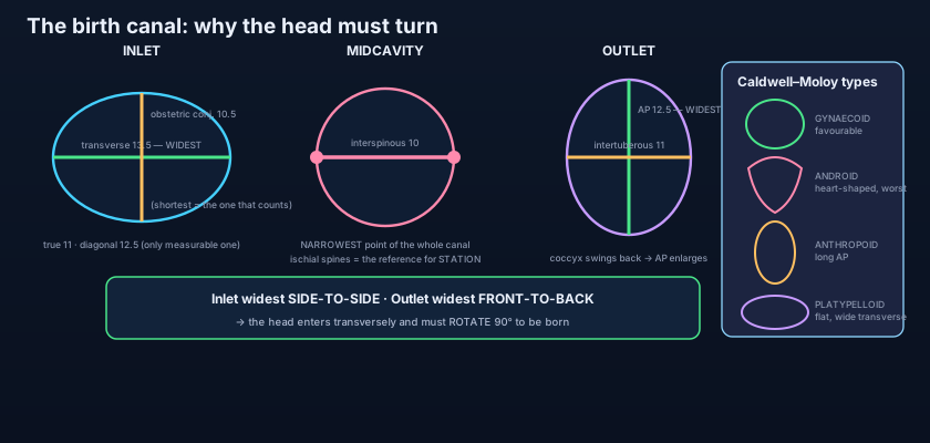
Three planes matter: the inlet, the midcavity and the outlet. Each has its own bottleneck. At the INLET the anteroposterior measurements are a family of three, and confusing them is the classic examination trap. The TRUE (anatomical) CONJUGATE runs from the sacral promontory to the UPPER border of the pubic symphysis and measures about 11 cm. The OBSTETRIC CONJUGATE runs from the promontory to the point on the back of the symphysis that projects furthest into the pelvis — a few millimetres below the upper border — and measures about 10.5 cm. It is the SHORTEST anteroposterior diameter of the inlet, and therefore the only one the fetal head actually has to negotiate; it is the diameter that counts. The catch is that neither of those can be measured on a living woman, because a finger cannot get behind the symphysis. What CAN be measured is the DIAGONAL CONJUGATE, from the lower border of the symphysis back to the promontory, about 12.5 cm — the one the midwife in our opening scene was reaching for. Subtract 1.5 to 2 cm from it and you have estimated the obstetric conjugate; and if the promontory cannot be reached at all with the examining fingers, the inlet is by definition generous. The TRANSVERSE diameter of the inlet, the widest distance across the brim, is about 13.5 cm, and the OBLIQUE diameters, running from each sacroiliac joint to the opposite iliopubic eminence, are about 12 cm. Note the shape this produces: at the inlet, WIDE FROM SIDE TO SIDE.
In the MIDCAVITY the picture reverses and tightens. The plane of least dimensions passes through the lower border of the symphysis, the ISCHIAL SPINES and the fourth to fifth sacral segment, and the INTERSPINOUS DIAMETER between the two ischial spines is about 10 cm. That is the NARROWEST measurement in the entire pelvic canal, and it is where an arrest of labour most often occurs. Prominent, inward-pointing spines can reduce it further. At the OUTLET, the transverse or INTERTUBEROUS diameter between the inner margins of the ischial tuberosities is about 11 cm, and the ANTEROPOSTERIOR diameter, from the lower border of the symphysis to the tip of the coccyx, is about 12.5 cm — and it is the one measurement in the pelvis that can lengthen during the event itself, because the coccyx is pushed back on its mobile sacrococcygeal joint. So the outlet is WIDE FROM FRONT TO BACK. Put the two facts side by side — inlet widest transversely, outlet widest anteroposteriorly — and the entire mechanism of labour falls out of the geometry: the head must ENGAGE transversely, DESCEND, and ROTATE ninety degrees at the level of the spines before it can be delivered. It is not a quirk of the fetus; it is the pelvis dictating terms. The sequence itself is followed step by step in the anatomy of childbirth.
Two bony points on the pelvic side wall carry more clinical weight than any other landmark in the region, and they are the ISCHIAL SPINES. First, they define the narrowest plane of the canal, so the level of the presenting part is measured against them: when the leading bony point of the fetal head is level with the spines the head is at STATION 0 and, by definition, engaged; above them stations are negative, below them positive, in centimetres. Second, the spine is where the pudendal nerve is at its most accessible: the nerve leaves the pelvis through the greater sciatic foramen, hooks around the back of the SACROSPINOUS LIGAMENT exactly at its attachment to the spine, and re-enters through the lesser sciatic foramen — so a needle passed along the examining finger to the spine, transvaginally, will find it. That is a PUDENDAL BLOCK, the anaesthetic for an instrumental delivery or a perineal repair, and it is described in the pudendal nerve. One landmark, two entirely different uses — which is why the examining finger goes looking for it every time.
Four shapes: the Caldwell–Moloy types
Pelvises are not interchangeable, and in 1933 Caldwell and Moloy sorted them into four types by the shape of the inlet. The GYNAECOID pelvis is the classic favourable female form and the commonest, found in roughly half of women: a rounded, slightly transversely oval inlet, straight side walls, a shallow non-prominent sacral promontory, blunt widely spaced ischial spines, a well-curved sacrum and a wide subpubic arch. Everything about it is generous, and it is the pelvis that delivers easily. The ANDROID pelvis is the typical male form, present in a minority of women, and it is the least favourable of the four: the inlet is HEART-SHAPED because the promontory juts forward and the forepelvis — the space in front of the transverse diameter — is narrow and wedge-like; the side walls converge downwards, the ischial spines are prominent, the sacrum is flat and inclined forwards, and the subpubic angle is narrow. A head that descends into it is squeezed backwards into a shrinking posterior segment, and obstructed labour, deep transverse arrest and difficult instrumental delivery are all commoner. The ANTHROPOID pelvis has an inlet that is a long OVAL in the anteroposterior direction — the anteroposterior diameter exceeds the transverse — with a long, narrow, deep cavity; the head often engages and stays in the occipito-posterior or direct occipito-anterior position, and delivery, though sometimes slow, is usually possible. The PLATYPELLOID (simple flat) pelvis is the rarest: the inlet is flattened, wide from side to side and short from front to back, so the head has trouble ENTERING at all and must engage with marked lateral tilt. In practice pelvises are mixed types, and the classification is best used as a way of thinking about which diameter is likely to be limiting.
Male and female, point by point
The pelvis is the most sexually dimorphic part of the human skeleton — and the differences are all obstetric in purpose. Take them one at a time. The SUBPUBIC ANGLE beneath the pubic arch is wide in the female, greater than 80–90° — classically about the angle between your thumb and index finger held apart — and narrow in the male, roughly 50–60°, about the angle between two adjacent fingers. The PELVIC INLET is rounded or transversely oval in the female and heart-shaped in the male, whose forward-jutting promontory intrudes on it. The ISCHIAL TUBEROSITIES are EVERTED (turned outwards) in the female, widening the intertuberous diameter, and inverted in the male. The GREATER SCIATIC NOTCH is wide and shallow in the female, approaching 60°, and narrow and deep in the male, nearer 40–50°. The ISCHIAL SPINES are blunt and turned outwards in the female, sharper and more inward-projecting in the male. The whole female pelvis is SHALLOWER and WIDER, with a broader, shorter sacrum that is less curved, thinner and lighter bones with less prominent muscle markings, and a larger, more circular obturator foramen in the male versus a smaller triangular one in the female. The ACETABULA are smaller and face more anteriorly in the female and lie further apart, which is why the femora converge more steeply to the knee. Because these differences are so consistent, the pelvis is the single most reliable bone in the skeleton for determining sex in forensic and archaeological work: a well-preserved pelvis will be sexed correctly far more often than a skull, and the greater sciatic notch and subpubic angle alone carry most of that accuracy.
The ring: joints, ligaments and the forces they fight
Three joints close the ring, and every one of them is built to be strong rather than mobile. Behind, the two SACROILIAC JOINTS transmit the entire weight of the head, arms and trunk from the vertebral column into the hip bones and onwards to the femora. Each is a hybrid: synovial in front between the auricular surfaces, and a syndesmosis behind, where the immensely strong INTEROSSEOUS SACROILIAC LIGAMENT fills the space between the rough tuberosities of sacrum and ilium and effectively SUSPENDS the sacrum from the iliac bones. Anterior and posterior sacroiliac ligaments reinforce it; the joint surfaces themselves are irregular and interlocking, and movement is limited to a few degrees of rocking (nutation and counter-nutation). In front, the PUBIC SYMPHYSIS is a SECONDARY CARTILAGINOUS JOINT (a symphysis): the two pubic bones are capped with hyaline cartilage and united by a fibrocartilaginous interpubic disc, reinforced above and below by the superior pubic and arcuate pubic ligaments. It is a strut, not a hinge, and it resists the medial compression squeezing the two halves together. In pregnancy, under RELAXIN and the sex steroids, the interpubic disc softens and the joint widens by several millimetres; the sacroiliac joints loosen in parallel. The gain is a slightly larger canal at delivery. The cost is instability, and when it becomes symptomatic — pain over the symphysis and groins, worse on walking, climbing stairs or turning in bed, sometimes with an audible click and a waddling gait — it is called symphysis pubis dysfunction, or pelvic girdle pain.
Now the elegant part. Stand up, and the weight of the trunk bears down on the sacrum in front of the sacroiliac joints. That force tends to rotate the sacrum about a transverse axis so that the PROMONTORY TIPS FORWARD AND DOWNWARDS while the lower end and the coccyx swing BACKWARDS AND UPWARDS. Nothing on the front of the joint can stop that rotation. What stops it are two accessory ligaments slung between the sacrum and the ischium: the SACROTUBEROUS LIGAMENT, running from the posterior iliac spines, lower sacrum and coccyx to the ischial tuberosity, and the SACROSPINOUS LIGAMENT, running from the lateral sacrum and coccyx to the ischial spine, lying deep to it. Both are attached to the LOWER end of the sacrum, and both anchor it downwards to the ischium — so as the promontory tries to tip forwards, they tighten on the far end of the lever and hold the whole bone still. They are, in effect, guy ropes on a tilting mast. And in doing that job they also convert the two notches of the hip bone into two openings: above the sacrospinous ligament, the GREATER SCIATIC FORAMEN, the exit route from the pelvis to the gluteal region for piriformis, the sciatic and gluteal nerves and vessels and the pudendal nerve; and between the two ligaments, the LESSER SCIATIC FORAMEN, the entrance to the perineum for the tendon of obturator internus, the pudendal nerve and the internal pudendal vessels. One mechanical solution, two anatomical doorways.
A woman in her first labour reaches full dilatation and pushes for two hours. The head is at station 0, wedged at the level of the ischial spines, and it does not rotate: the sagittal suture stays transverse. This is DEEP TRANSVERSE ARREST, and on examination the reason is written in the bone — a prominent, forward-jutting promontory, converging side walls, sharp spines that can be felt easily, and a subpubic arch that admits barely one finger. This is an android pelvis, and its narrow forepelvis leaves no room in front for the head to turn into. The head cannot enter the anterior segment, cannot rotate, and cannot descend; the outlet, narrow between inverted tuberosities, offers nothing better below. This is CEPHALOPELVIC DISPROPORTION — not because the baby is unusually large, but because the passage is the wrong shape. It is diagnosed by failure to progress despite adequate contractions, not by measurement in advance, because pelvimetry predicts labour poorly. The answer is a caesarean section, and the anatomy of the failure is the reason the operation exists. The uterine side of the same problem — the contractions, their augmentation and their limits — belongs to labour induction and augmentation.
- The human pelvis is a compromise in the OBSTETRIC DILEMMA: efficient bipedal walking favours a narrow pelvis, a large-brained newborn demands a wide one — and almost every feature of the cavity is a negotiation between the two.
- The GREATER (false) pelvis lies above the brim and is functionally abdomen; the LESSER (true) pelvis lies below it and is the pelvic cavity proper — anterior wall only about 4 cm, posterior wall about 12 cm, which is why the canal is curved.
- The PELVIC BRIM (inlet) is traced: sacral promontory → anterior ala of sacrum → arcuate line of ilium → pecten pubis (pectineal line) → pubic crest → upper border of the pubic symphysis.
- The OUTLET is a diamond: pubic arch in front, ischial tuberosities at the sides, sacrotuberous ligaments and the coccyx behind — the back of it is ligament and mobile coccyx, so it can lengthen at delivery.
- The pelvic AXIS (curve of Carus) runs down and back, then turns forwards at the level of the ischial spines: the fetus negotiates a bend, not a straight tube — which is why forceps have a pelvic curve.
- The pelvic inlet is tilted about 55–60° to the horizontal in the anatomical position, so the anterior superior iliac spine and the pubic tubercle lie in one vertical plane.
- INLET diameters: true (anatomical) conjugate ~11 cm; OBSTETRIC conjugate ~10.5 cm (shortest AP, the one that counts, not measurable clinically); DIAGONAL conjugate ~12.5 cm (the only one measurable — subtract 1.5–2 cm to estimate the obstetric); TRANSVERSE ~13.5 cm (widest at the inlet).
- MIDCAVITY: the INTERSPINOUS diameter between the ischial spines is ~10 cm — the NARROWEST point in the whole canal, and the reference level for fetal STATION (station 0 = engaged).
- OUTLET: intertuberous (transverse) ~11 cm; anteroposterior ~12.5 cm, and it ENLARGES as the coccyx is pushed back. Inlet widest TRANSVERSELY + outlet widest ANTEROPOSTERIORLY = the head must rotate 90° as it descends.
- CALDWELL–MOLOY types: GYNAECOID (rounded, favourable, commonest), ANDROID (heart-shaped, narrow forepelvis, prominent spines — least favourable), ANTHROPOID (long AP oval), PLATYPELLOID (flat: wide transverse, short AP — rarest).
- SEXUAL DIMORPHISM: female subpubic angle >80–90° (male ~50–60°), rounded versus heart-shaped inlet, everted ischial tuberosities, wider and shallower greater sciatic notch (~60° vs ~40–50°), blunter outward-turned spines, shallower and wider pelvis with lighter bones. The pelvis is the most reliable bone for forensic sex determination.
- The RING: two sacroiliac joints (weight-transmitting; the interosseous sacroiliac ligament suspends the sacrum), the pubic symphysis (a secondary cartilaginous joint that softens under relaxin in pregnancy), and the sacrotuberous and sacrospinous ligaments, which resist forward tipping of the promontory and convert the sciatic notches into the greater and lesser sciatic foramina.
- Confusing the three inlet conjugates. The TRUE conjugate (~11 cm) goes to the UPPER border of the symphysis; the OBSTETRIC conjugate (~10.5 cm) is the SHORTEST and the one the head must pass, but it cannot be measured in life; the DIAGONAL conjugate (~12.5 cm) is the only one a finger can reach, and the obstetric is estimated from it by subtracting 1.5–2 cm.
- Assuming the inlet is the tightest part of the canal. It is not: the narrowest measurement in the whole pelvis is the INTERSPINOUS diameter (~10 cm) at the midcavity, which is why arrest of descent so often happens at the level of the ischial spines.
- Reading a single displaced fracture of the pelvic ring as an isolated injury. A rigid ring cannot displace at one point only — find the second break, and remember the posterior (sacroiliac) disruption is the one that bleeds, with the binder applied at the greater trochanters rather than the iliac crests.
A woman in labour reaches full dilatation, but after two hours of pushing the fetal head remains level with the ischial spines with its sagittal suture lying transversely. Which diameter is the head failing to negotiate, and what is its approximate normal value?
- The pelvis is a compromise in the obstetric dilemma. The brim divides a GREATER (false) pelvis above — functionally abdomen — from the LESSER (true) pelvis below, the real pelvic cavity, whose 4 cm anterior and 12 cm posterior walls make the canal a curve (the axis of Carus) rather than a tube.
- The brim runs promontory → arcuate line → pecten pubis → pubic crest → upper symphysis; the outlet is a diamond of pubic arch, ischial tuberosities, sacrotuberous ligaments and coccyx, and it lengthens as the coccyx swings back.
- Diameters: inlet — true conjugate ~11, OBSTETRIC ~10.5 (shortest, decisive, unmeasurable), DIAGONAL ~12.5 (the only clinical one), transverse ~13.5; midcavity — INTERSPINOUS ~10, the narrowest point and the reference for station; outlet — intertuberous ~11, anteroposterior ~12.5. Inlet widest transversely + outlet widest anteroposteriorly = the head must rotate 90°.
- Four Caldwell–Moloy types (gynaecoid, android, anthropoid, platypelloid); a wide subpubic angle, everted tuberosities and a wide sciatic notch mark the female pelvis (the best forensic sex indicator); and the ring is held by two sacroiliac joints, the pubic symphysis (relaxin-softened in pregnancy) and the sacrotuberous and sacrospinous ligaments, which resist forward tipping of the promontory and create the greater and lesser sciatic foramina.
- Drake RL, Vogl AW, Mitchell AWM. Gray's Anatomy for Students — Pelvis and Perineum: the pelvic girdle, pelvic inlet and outlet, and pelvic measurements.
- Moore KL, Dalley AF, Agur AMR. Clinically Oriented Anatomy — The bony pelvis, sexual differences, pelvic diameters and the obstetric significance of the pelvic cavity.
- Netter FH. Atlas of Human Anatomy — Bony framework of the pelvis; male and female pelvis compared; pelvic diameters.
- Last RJ. Last's Anatomy: Regional and Applied — The pelvic girdle, sacroiliac joints, sacrotuberous and sacrospinous ligaments.
- Snell RS. Clinical Anatomy by Regions — The pelvis: false and true pelvis, pelvic types, and clinical notes on pelvic fractures and labour.
- Caldwell WE, Moloy HC. Anatomical variations in the female pelvis and their effect in labor. American Journal of Obstetrics and Gynecology.

