
The Thyroid: The Gland That Moves When You Swallow
Put a finger on the front of your neck, just below the Adam's apple, and swallow. Something rises under your fingertip and settles back — a soft butterfly of tissue that is bound so tightly to the windpipe it has no choice but to travel with it. That single movement is the oldest bedside sign in endocrine surgery: a lump that rides up when you swallow is thyroid; a lump that sits still is not. But the same gland that betrays itself so obligingly hides a danger. It is wrapped around two nerves that a surgeon must find and spare, drinks from arteries fed by the biggest vessels in the neck, and shelters on its back four pinhead glands that rule the calcium in your blood. Cut carelessly, and a patient wakes hoarse, or unable to breathe, or with hands that cramp into spasm. This is the anatomy of a gland that everyone can feel and few can operate on safely.
A 40-year-old woman comes to clinic because a friend noticed a swelling low in the front of her neck. She has felt well. The examiner does not reach for a scanner first — he reaches for a glass of water. He stands behind her, rests the pads of his fingers over the lower neck, and asks her to take a mouthful and swallow. Under his fingers a firm, symmetrical mass glides upward two centimetres and drops back. Then he asks her to poke her tongue out as far as it will go: the mass does not budge. Two questions of physiology, answered with a sip of water and a stuck-out tongue, have already told him more than a photograph could. The upward glide on swallowing says the mass is bound to the larynx and trachea inside the pretracheal fascia — it is thyroid, or moves with the thyroid. The stillness on tongue protrusion says it is NOT tethered to the base of the tongue by a thyroglossal remnant — so it is a goitre, not a thyroglossal cyst. He has localised the lump to a single gland before laying a finger flat on it, because he understands why this gland, alone among the lumps of the neck, must move when you swallow.
A butterfly wrapped round the windpipe
Two lobes, one bridge, and — in many people — a stray finger of tissue pointing upward. The thyroid gland is shaped like a butterfly, or a bow tie. It has two lateral LOBES, each pressed against the side of the larynx and upper trachea, joined across the midline by a narrow bridge, the ISTHMUS, which lies across the SECOND, THIRD and FOURTH tracheal rings. In perhaps a third to a half of people a slender PYRAMIDAL LOBE runs upward from the isthmus (usually a little to the left) towards the hyoid bone — a leftover marker of the path the gland travelled during development, and a piece of tissue the surgeon must remember to remove in a total thyroidectomy or it will light up on a later scan. Each lobe has intimate neighbours that decide everything a surgeon does. LATERALLY, the lobe is moulded against the CAROTID SHEATH with the common carotid artery, the internal jugular vein and the vagus nerve running inside it — the great vessels charted in the carotid and jugular. MEDIALLY, it embraces the larynx, the trachea, the pharynx and the oesophagus. And POSTERIORLY, on the back of the lobe, sit the parathyroid glands and, in the groove between trachea and oesophagus, the nerve that dominates this whole story.
Why it moves when you swallow
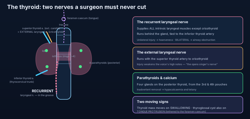
The whole gland is wrapped in a sleeve of PRETRACHEAL FASCIA, the layer of deep cervical fascia that also invests the larynx and trachea. This is the key to the swallowing sign. Because the fascia binds the thyroid to the front of the larynx and trachea, and because the larynx is hauled upward every time you swallow, the thyroid has no choice but to ride up with it and slide back down. A lump that is truly part of, or fixed to, this fascial envelope MOVES ON SWALLOWING; a lymph node, a skin cyst, or a lipoma sitting outside the envelope does not. That one observation separates a thyroid swelling from almost every other lump in the front of the neck — the systematic approach to which is laid out in the neck lump. Note the deliberate contrast the examiner draws with the tongue: a THYROGLOSSAL CYST, which is anchored to the base of the tongue through the tract the gland descended along, rises specifically on tongue PROTRUSION, whereas the thyroid rises on SWALLOWING. Two swellings, two movements, two different tethers — and both are simply anatomy made visible at the bedside.
The gland that came down from the tongue
Every thyroid anomaly is a story about a journey that did not go to plan. The thyroid does not begin in the neck. It begins in the floor of the developing pharynx, at a spot on the back of the tongue that you can still see in the adult as a small pit — the FORAMEN CAECUM. From there the primordial gland descends through the tissues of the neck, in front of the hyoid and the laryngeal cartilages, trailing behind it a narrow channel: the THYROGLOSSAL DUCT. Normally the duct disappears completely once the gland reaches its final home over the trachea, and the foramen caecum is all that remains. When the duct does NOT fully involute, its remnants explain a family of midline anomalies. A persisting pocket of duct becomes a THYROGLOSSAL CYST — a smooth, midline swelling, classically at or near the hyoid, that rises on tongue protrusion because it is still tethered up to the foramen caecum. If the gland fails to descend at all, thyroid tissue is left high up as a LINGUAL THYROID at the back of the tongue, which may be the person's ONLY thyroid tissue — a crucial thing to know before anyone removes it. And the pyramidal lobe seen at operation is simply the lowest, persistent part of that same duct. The migration also shares its territory with the pharyngeal pouches that give rise to the parathyroids and thymus, a developmental neighbourhood set out in the pharyngeal arches.
A blood supply rich enough to be dangerous
Two arteries from two different sources, each running with a nerve you must not cut. For its size the thyroid is one of the most vascular organs in the body, and that richness is the surgeon's chief problem. Two paired arteries feed it. The SUPERIOR THYROID ARTERY is the FIRST branch of the EXTERNAL CAROTID ARTERY; it descends to the UPPER POLE of each lobe, and as it does so it runs in dangerously close company with the EXTERNAL BRANCH OF THE SUPERIOR LARYNGEAL NERVE. The INFERIOR THYROID ARTERY arises from the THYROCERVICAL TRUNK of the subclavian artery, arches up and medially behind the carotid sheath, and reaches the LOWER POLE, where it crosses — and is intimately entangled with — the RECURRENT LARYNGEAL NERVE. There is, in some people, a small unpaired THYROIDEA IMA artery running up to the isthmus from the brachiocephalic trunk or the arch of the aorta, a hazard in a low tracheostomy. The venous drainage is by three routes: the SUPERIOR and MIDDLE thyroid veins empty into the INTERNAL JUGULAR VEIN, while the INFERIOR thyroid veins run down in front of the trachea to drain into the BRACHIOCEPHALIC VEINS. The surgical rule that falls out of this arrangement is simple and life-saving: tie the SUPERIOR artery low and close to the gland to stay away from the external laryngeal nerve, and tie the INFERIOR artery well away from the gland after positively identifying the recurrent laryngeal nerve.
Think of the thyroid as a house being demolished, with two live power cables running through the walls — one along the top of the building, one across the basement floor. You cannot pull the house down until the power is cut, and the power is cut by clamping the water and gas that feed it (the arteries). But each supply pipe is strapped to a live cable along its whole length. Grab the upper pipe carelessly and you crush the cable that dims the lights (the external laryngeal nerve — the voice loses its top notes). Grab the lower pipe carelessly and you cut the cable that runs the whole house (the recurrent laryngeal nerve — the voice fails, and if both go, the doors that let you breathe swing shut). The safe demolisher does the opposite of what haste suggests: he does not avoid the cables by working fast and far from them. He finds each cable first, follows it with his eyes, and only then cuts the pipe — hugging the gland where the upper cable has already peeled away, staying wide of the gland where the lower cable can be seen and protected.
The two nerves the surgeon dreads
This is the spine of the whole operation. The RECURRENT LARYNGEAL NERVE, a branch of the vagus, supplies ALL the intrinsic muscles of the larynx EXCEPT the cricothyroid, and it ascends to the larynx in the TRACHEO-OESOPHAGEAL GROOVE, running just behind the thyroid lobe and looped around, or embedded beside, the inferior thyroid artery. Its function and the movements of the vocal folds it controls belong to the larynx; what matters here is its fragility. UNILATERAL injury paralyses one vocal fold and leaves the patient HOARSE, often with a weak, breathy voice. BILATERAL injury is far worse: both folds drift towards the midline and cannot abduct, and the result is AIRWAY OBSTRUCTION — stridor after extubation, sometimes an emergency requiring re-intubation or tracheostomy. The nerve must therefore be actively identified and preserved, not merely avoided. Its course is asymmetrical because of the way the great vessels formed in the embryo: on the LEFT it hooks under the arch of the aorta, on the RIGHT under the right subclavian artery, so the right nerve reaches the groove more obliquely — and in a small number of people the right nerve is "NON-RECURRENT," branching straight off the vagus in the neck (a variant tied to an anomalous subclavian artery), a trap that has cost many a voice. That long thoracic detour, and why the two sides differ, belongs to the nerves of the thorax. The second nerve is the EXTERNAL BRANCH OF THE SUPERIOR LARYNGEAL NERVE, which runs with the superior thyroid artery to supply the CRICOTHYROID — the muscle that tenses the vocal fold. Injuring it does not obstruct the airway or cause obvious hoarseness; it robs the voice of its ability to reach and project high notes, which is why it is nicknamed the "opera singer's nerve" and why singers are counselled specifically about it before thyroid surgery.
The four glands on the back of the thyroid
Pinhead organs that rule the calcium in your blood — and are lost with a single careless snip. Behind the thyroid, usually four in number, lie the PARATHYROID GLANDS — each no bigger than a grain of rice, yellow-brown, embedded on the POSTERIOR surface of the thyroid lobes just under its capsule. They are the master regulators of CALCIUM: their PARATHYROID HORMONE (PTH) raises blood calcium by acting on bone, the kidney and (via vitamin D) the gut. Their embryology explains where they hide. The SUPERIOR pair derive from the FOURTH pharyngeal pouch; they travel only a short distance, so they are relatively CONSTANT in position, typically found at the level of the middle of the thyroid lobe near where the recurrent laryngeal nerve meets the inferior thyroid artery. The INFERIOR pair derive from the THIRD pharyngeal pouch and — counter-intuitively — migrate DOWN alongside the descending THYMUS (also a third-pouch derivative). Because they hitch a ride on the thymus, the inferior parathyroids are far more VARIABLE: they may sit at the lower pole, or be dragged right down into the superior MEDIASTINUM with thymic tissue. Their blood supply is mainly from the INFERIOR THYROID ARTERY (for both pairs), which is why that vessel must be ligated distally, close to the thyroid, sparing the tiny branches to the parathyroids. The clinical consequence is one of the most feared complications of thyroid surgery: inadvertent removal, bruising or DEVASCULARISATION of the parathyroids causes acute HYPOCALCAEMIA, and the patient develops perioral tingling, tingling fingers, and TETANY — Trousseau's and Chvostek's signs, and in severe cases carpopedal spasm or laryngospasm. It is why every thyroidectomy patient has their calcium watched, and why a good surgeon reimplants a parathyroid he cannot save.
One gland, but two completely different hormonal systems live inside it — and this is a favourite point of confusion. The THYROID follicular cells make the thyroid hormones T4 and T3, which set the body's metabolic rate; their overactivity and underactivity are treated in antithyroid drugs and radioiodine and levothyroxine replacement. Scattered between the follicles are the PARAFOLLICULAR (C) CELLS, which make CALCITONIN, a hormone that lowers calcium (and is a tumour marker for medullary thyroid carcinoma). And behind the gland entirely, on separate little organs, the PARATHYROIDS make PTH, which RAISES calcium. So calcium is regulated by a push-pull pair whose two halves sit millimetres apart but are embryologically and functionally distinct: calcitonin from inside the thyroid pushes calcium down, PTH from the parathyroids behind it pulls calcium up — and of the two, it is PTH that truly matters, which is exactly why losing the four little glands is so much more dangerous than losing the big one.
- The thyroid has two LOBES joined by an ISTHMUS over the 2nd–4th tracheal rings, ± a PYRAMIDAL LOBE (thyroglossal remnant) running up from the isthmus. It is enclosed in PRETRACHEAL FASCIA, which binds it to the larynx/trachea — hence it MOVES ON SWALLOWING.
- Relations of each lobe: LATERAL = carotid sheath (common carotid, IJV, vagus); MEDIAL = larynx, trachea, pharynx, oesophagus; POSTERIOR = parathyroids and the recurrent laryngeal nerve in the tracheo-oesophageal groove.
- Embryology: the gland arises at the FORAMEN CAECUM of the tongue and descends along the THYROGLOSSAL DUCT. A persistent duct → a midline THYROGLOSSAL CYST that rises on TONGUE PROTRUSION; failed descent → a LINGUAL/ectopic thyroid.
- Arteries: SUPERIOR THYROID (first branch of the EXTERNAL CAROTID) to the upper pole with the EXTERNAL LARYNGEAL NERVE; INFERIOR THYROID (from the THYROCERVICAL TRUNK) to the lower pole with the RECURRENT LARYNGEAL NERVE. Veins: superior + middle → IJV; inferior → brachiocephalic veins.
- The RECURRENT LARYNGEAL NERVE supplies all intrinsic laryngeal muscles except cricothyroid: UNILATERAL injury → hoarseness; BILATERAL injury → airway obstruction. The EXTERNAL LARYNGEAL NERVE (with the superior thyroid artery) supplies cricothyroid — injury weakens high notes.
What the gland makes, and where it drains
The thyroid's follicular cells trap iodide and manufacture THYROXINE (T4) and TRIIODOTHYRONINE (T3), storing them uniquely OUTSIDE the cell in the colloid of the follicle until called upon — the hormones that set metabolic rate, and whose excess or deficiency is the everyday business of the endocrine clinic. Its parafollicular C cells add CALCITONIN, and the parathyroids behind it add PTH, completing the calcium axis. The LYMPHATIC drainage matters because it is the map along which thyroid cancer spreads. Lymph runs from the isthmus and the medial upper poles to the PRETRACHEAL (DELPHIAN) node just above the isthmus — a node whose enlargement is a classic early warning — and from the rest of the gland to the PARATRACHEAL nodes in the tracheo-oesophageal grooves (the central, level VI compartment) and then to the DEEP CERVICAL chain along the internal jugular vein. This is why a thyroid cancer can present not as a lump in the gland but as a hard node low in the neck, and why the surgeon planning a cancer operation thinks in terms of these nodal levels, not just the gland itself.
A 55-year-old woman has a total thyroidectomy for a multinodular goitre. The operation is uneventful. In recovery she is comfortable, but when she speaks her voice is weak and breathy, and she tires after a few sentences. Flexible laryngoscopy shows the LEFT vocal fold lying still in a paramedian position: a UNILATERAL recurrent laryngeal nerve palsy, most likely a stretch or bruise rather than a division, since the nerve was seen intact at operation. She is reassured that many such neuropraxias recover over weeks to months, and offered speech therapy; the healthy right fold will often compensate by crossing the midline. Now imagine the far graver alternative: had BOTH recurrent laryngeal nerves been injured, both folds would sit near the midline and be unable to abduct, and the danger would not be the voice but the AIRWAY — stridor on extubation, and a real possibility of emergency re-intubation or tracheostomy. This is why the modern operation identifies each nerve before dividing anything near it, often with an intra-operative nerve monitor, and why a surgeon documents vocal fold movement before and after. Separately, if this same patient had gone home and returned two days later with tingling lips and fingers and cramping hands, the diagnosis would be HYPOCALCAEMIA from bruised or devascularised PARATHYROIDS — the other price of operating in this crowded corner of the neck.
- The PARATHYROIDS (usually four) sit on the POSTERIOR surface of the thyroid lobes and secrete PTH, the master regulator of calcium. The SUPERIOR pair (4th pouch) are constant; the INFERIOR pair (3rd pouch) migrate with the THYMUS and are variable, sometimes reaching the mediastinum. Both are supplied mainly by the INFERIOR THYROID ARTERY.
- Inadvertent removal or DEVASCULARISATION of the parathyroids in thyroid surgery causes HYPOCALCAEMIA and TETANY (perioral/finger tingling, Chvostek's and Trousseau's signs, carpopedal spasm) — a chief reason to ligate the inferior thyroid artery close to the gland.
- Two hormone systems: THYROID follicular cells make T4/T3 (metabolic rate); PARAFOLLICULAR (C) cells make CALCITONIN (lowers calcium); the parathyroids make PTH (raises calcium). PTH is the one that truly matters clinically.
- LYMPHATIC drainage: to the pretracheal (DELPHIAN) node above the isthmus, the paratracheal (central, level VI) nodes, and the deep cervical chain — the routes along which thyroid cancer spreads, so cancer may present as a hard low neck node.
- The MOVING-ON-SWALLOWING sign (pretracheal fascia) distinguishes a thyroid mass from other neck lumps; a THYROGLOSSAL CYST instead moves on TONGUE PROTRUSION; a RETROSTERNAL goitre can extend behind the sternum and cause tracheal compression.
- Confusing the two nerves and their consequences. The RECURRENT laryngeal nerve (with the INFERIOR thyroid artery) supplies all intrinsic laryngeal muscles except cricothyroid — its injury causes hoarseness (unilateral) or airway obstruction (bilateral). The EXTERNAL laryngeal nerve (with the SUPERIOR thyroid artery) supplies only cricothyroid — its injury just weakens the high notes of the voice.
- Thinking a thyroglossal cyst and a thyroid goitre move the same way. A goitre (bound in the pretracheal fascia) moves on SWALLOWING; a thyroglossal cyst (tethered up to the foramen caecum) moves specifically on TONGUE PROTRUSION. And never remove a lingual/ectopic thyroid before confirming there is normal thyroid tissue elsewhere — it may be the only functioning thyroid the patient has.
- Forgetting the parathyroids when operating on the thyroid. Ligating the inferior thyroid artery far from the gland, or stripping the posterior capsule, devascularises the parathyroids and causes hypocalcaemia — the artery must be taken distally, close to the thyroid, preserving its tiny parathyroid branches; a parathyroid that cannot be saved should be reimplanted.
During a total thyroidectomy the surgeon is ligating the vessels at the LOWER pole of the thyroid lobe. Which nerve is most closely related to the artery being tied here, and what is the consequence of injuring it bilaterally?
- The thyroid is a butterfly of two LOBES joined by an ISTHMUS over the 2nd–4th tracheal rings (± a PYRAMIDAL LOBE), wrapped in PRETRACHEAL FASCIA that binds it to the larynx and trachea — so it MOVES ON SWALLOWING, the sign that identifies a thyroid mass; a THYROGLOSSAL CYST instead moves on TONGUE PROTRUSION.
- It develops at the FORAMEN CAECUM of the tongue and descends along the THYROGLOSSAL DUCT; failure of this to disappear gives a midline thyroglossal cyst, a lingual/ectopic thyroid or the pyramidal lobe. It shares its developmental territory with the pharyngeal pouches that form the parathyroids and thymus.
- Blood supply is rich and dangerous: the SUPERIOR THYROID artery (first branch of the EXTERNAL CAROTID) runs to the upper pole with the EXTERNAL LARYNGEAL NERVE (high notes), and the INFERIOR THYROID artery (from the THYROCERVICAL TRUNK) to the lower pole with the RECURRENT LARYNGEAL NERVE (tracheo-oesophageal groove). Veins: superior/middle → IJV, inferior → brachiocephalic.
- The recurrent laryngeal nerve supplies all intrinsic laryngeal muscles except cricothyroid — unilateral injury → hoarseness, bilateral → airway obstruction; both nerves must be identified and preserved. Behind the gland the four PARATHYROIDS (superior = 4th pouch, constant; inferior = 3rd pouch with the thymus, variable) secrete PTH and rule calcium — their loss in surgery causes HYPOCALCAEMIA and tetany, and links to the pharmacology of hyper- and hypothyroidism.
- Drake RL, Vogl AW, Mitchell AWM. Gray's Anatomy for Students — Neck: the thyroid and parathyroid glands, their arteries and the laryngeal nerves.
- Moore KL, Dalley AF, Agur AMR. Clinically Oriented Anatomy — The thyroid gland, thyroidectomy and the recurrent and external laryngeal nerves.
- Netter FH. Atlas of Human Anatomy — Thyroid gland and pharynx; arteries and nerves of the thyroid region.
- Last RJ. Last's Anatomy: Regional and Applied — The thyroid and parathyroid glands and the tracheo-oesophageal groove.
- Snell RS. Clinical Anatomy by Regions — The thyroid gland, the thyroglossal duct and thyroid swellings.
- TeachMeAnatomy — The Thyroid Gland; The Parathyroid Glands.

