
The Skull: A Box Built From Twenty-Two Bones
The skull is the most complicated bone in the body, and it is not one bone at all but twenty-two, locked together along jagged seams into a single rigid box. That box has two jobs it never sets down: to carry the face, and to protect the brain. To do the second it is thick and strong almost everywhere — except at one coin-sized patch above the ear, where four bones meet over the trunk of an artery, and where a single unlucky blow can start a bleed that kills a person who was, an hour before, walking and talking and feeling fine. And the floor of the box is not flat. It steps down in three broad terraces, each cradling a different part of the brain, each pierced by holes that let the nerves and vessels in and out — and each, for that reason, a line along which the base will crack when the head is struck hard enough. To read a skull is to read both an engineering solution and a map of where things go wrong.
A twenty-year-old is brought in after a fight outside a bar. He took a single punch to the side of the head, above and in front of the ear, went down, was briefly stunned, then got up, argued with the doorman, and walked to the taxi rank on his own two feet. His friends say he seemed completely normal — talking, joking, a little embarrassed. That stretch of ordinary behaviour has a name that every clinician learns to fear: the LUCID INTERVAL. An hour later he is drowsy. By the time he reaches the department he is barely rousable, his right pupil is blowing wide and fixed, and his left arm is not moving. The CT shows a lens-shaped, biconvex pool of fresh blood pressed against the inner surface of the skull, exactly under that spot above the ear. The bone there is called the pterion, and it is the thinnest part of the entire vault. The blow cracked it, the crack tore the artery running just beneath, and the artery — not a slow vein but a pressurised artery — has been filling the space between bone and brain-covering ever since, quietly, through the very minutes he spent seeming fine.
Twenty-two bones, eight of them around the brain
Count them once and the architecture stops being a list and becomes a building. The skull is assembled from TWENTY-TWO bones: EIGHT CRANIAL bones that enclose the brain, and FOURTEEN FACIAL bones that build the face beneath it. The cranial eight are the FRONTAL bone in front, the two PARIETAL bones forming the roof and sides, the OCCIPITAL bone behind and below, the two TEMPORAL bones low on each side, the single ETHMOID deep between the orbits, and the butterfly-shaped SPHENOID spanning the width of the base like a keystone. Almost all of them are FLAT bones of the kind set out in bones and their structure — formed by intramembranous ossification, built of two hard plates with spongy bone between — and they meet one another not at movable joints but at SUTURES, immovable fibrous joints where interlocking bony teeth are laced together, rigid in the adult and unmistakable to the eye as jagged running lines. The whole assembly divides naturally into two: the domed CALVARIA or vault that caps the brain, and the CRANIAL BASE on which the brain sits. Take a skull apart along its sutures and it is a three-dimensional jigsaw; leave it whole and it is a single rigid shell, which is precisely the point.
The vault, its seams, and two named crossings
Three great sutures divide the roof, and where they meet, the bones are named after the gap that once lay there. Look down on the top of the vault and three sutures organise the whole roof. The CORONAL suture runs across the crown from side to side, separating the frontal bone in front from the two parietals behind. The SAGITTAL suture runs front-to-back along the midline between the two parietal bones. The LAMBDOID suture runs across the back, separating the parietals from the occipital bone, and named for its resemblance to the Greek letter lambda. Lower on each side the SQUAMOUS suture arcs above the ear where the flat squamous part of the temporal bone overlaps the parietal like the edge of a scale. Two crossings have their own names and are landmarks a surgeon and a radiologist both use. The BREGMA is the point where the coronal and sagittal sutures meet — the junction of frontal and parietals — a soft gap in the newborn, a hard cross in the adult. The LAMBDA is the point where the sagittal and lambdoid sutures meet — the junction of parietals and occipital. In life these are not decoration: they are the reference points from which the surgeon measures, and the lines along which the infant skull can still move.
The pterion — a coin of thin bone over an artery
One H-shaped meeting of four bones is the single most dangerous point on the vault. On the side of the head, a couple of finger-breadths above the midpoint of the zygomatic arch, four bones come together in a small H-shaped suture pattern: the FRONTAL, the PARIETAL, the SQUAMOUS part of the TEMPORAL, and the GREATER WING of the SPHENOID. This meeting-place is the PTERION, and it is the THINNEST part of the skull — a place where four bones taper to their edges over a single small area. That would be merely a curiosity except for what lies on the inside surface directly beneath it: the ANTERIOR DIVISION of the MIDDLE MENINGEAL ARTERY, grooving the bone as it climbs. A blow to the temple that fractures the pterion tears that artery, and because it is an artery under systemic pressure, blood is driven into the space between the bone and the tough outer covering of the brain — stripping the DURA off the skull and forming an EXTRADURAL (epidural) HAEMATOMA, described with the coverings of the brain in the meninges and dural venous sinuses. On CT it is a BICONVEX (lens-shaped) collection that does not cross the sutures, because the dura is firmly anchored at the suture lines and the expanding blood cannot pass them. This is the lesion behind the boxer felled by a temple shot, the child struck by a cricket ball, and the young man of our opening scene — and the lucid interval is its signature, the deceptive quiet while the clot silently grows.
Think of the vault as the shell of a hard hat, moulded in one smooth curve, thick enough to take a knock almost anywhere. Now imagine that at one small spot on the side the manufacturer glued four separate offcuts together, edge to edge, and thinned them all down to save weight — and that immediately behind that patch, on the inside, they routed a pressurised water pipe. Everywhere else the hat can be struck without consequence. But a knock on that one patch does two things at once: it cracks the weakest part of the shell, and it splits the pipe behind it, so that water begins forcing its way in behind the lining while the wearer, feeling only a bump, carries on. That is the pterion exactly. The skull's strength is real but not uniform, and the body's misfortune is that its thinnest bone sits over one of the few arteries that runs between bone and dura. Nature did not put the artery there to be dangerous; it put the bone there thin, and the two happen to overlap.
The diploë, and the newborn's soft spots
Slice through a vault bone and you find it is not solid: two dense plates — an OUTER TABLE and an INNER TABLE of compact bone — sandwich a layer of spongy bone called the DIPLOË, its marrow spaces drained by wide, thin-walled DIPLOIC VEINS that connect the veins of the scalp outside to the dural venous sinuses inside. This tripled construction gives strength for little weight, but the inner table is thinner and more brittle than the outer, which is why a blow can fracture the inner table while the outer looks intact. In the NEWBORN the story is different again, because the sutures have not yet closed. Where several bones approach at the vault's corners there remain wide membranous gaps — the FONTANELLES. The ANTERIOR fontanelle, a diamond at the bregma between the two frontal and two parietal bones, is the largest and the last to close, at around EIGHTEEN MONTHS. The POSTERIOR fontanelle at the lambda closes far earlier, by two to three months, and two smaller LATERAL fontanelles — the sphenoidal and mastoid — sit low on each side. These gaps are not a weakness of design but a requirement of birth: they let the bones of the vault overlap and slide during delivery, MOULDING the head to pass through the pelvis, and afterwards they allow the vault to keep pace with a brain that doubles in size in the first year.
The base from inside — three terraced fossae
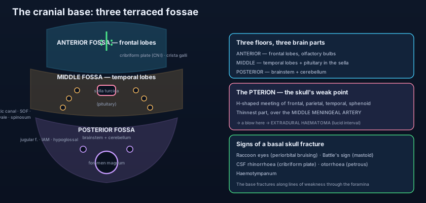
Lift off the vault and the floor of the box drops in three broad steps, each holding a different part of the brain. Seen from inside, the cranial base is not flat but TERRACED, stepping downwards from front to back in three FOSSAE. The ANTERIOR CRANIAL FOSSA is the highest and holds the FRONTAL LOBES. Its floor is built from the orbital plates of the FRONTAL bone, the CRIBRIFORM PLATE of the ETHMOID in the midline, and the LESSER WINGS of the SPHENOID behind. Rising from the cribriform plate is a small vertical blade, the CRISTA GALLI — literally the cock's comb — to which the falx cerebri anchors, and on either side of it lie the OLFACTORY BULBS, receiving the threads of the first cranial nerve as they pass up through the sieve-like holes of the cribriform plate from the nose, the nasal roof being explored in the nose and paranasal sinuses. The MIDDLE CRANIAL FOSSA steps down behind, deeper on each side and holding the TEMPORAL LOBES, its central part carrying the PITUITARY gland. Its floor is the butterfly of the SPHENOID — the central body with the GREATER WINGS spreading laterally — together with the TEMPORAL bones. And behind and below everything is the deep POSTERIOR CRANIAL FOSSA, the largest and lowest, cradling the BRAINSTEM and the CEREBELLUM, floored by the OCCIPITAL bone and the PETROUS parts of the temporal bones. Each step down matches the part of the brain it carries: the light frontal lobes ride highest, the great mass of the cerebellum sits deepest.
The sella turcica, and the holes in the floor
At the very centre of the base, on the body of the sphenoid, is a saddle-shaped hollow — the SELLA TURCICA, the "Turkish saddle" — and seated in its deepest part, the hypophyseal fossa, is the PITUITARY GLAND, the master endocrine gland, hanging on its stalk from the base of the brain and roofed by a fold of dura. Its position matters clinically because it sits directly below the OPTIC CHIASM: a pituitary tumour enlarging upward presses on the crossing fibres and classically robs a person of their outer fields of vision, a bitemporal hemianopia. The middle fossa is also the most heavily perforated part of the base. Through the SPHENOID pass, in order, the OPTIC CANAL (carrying the optic nerve into the orbit), the SUPERIOR ORBITAL FISSURE (the great slit into the orbit for the nerves of eye movement), the FORAMEN ROTUNDUM, the FORAMEN OVALE and the FORAMEN SPINOSUM. Each transmits a specific nerve or vessel, and their exact contents — which nerve leaves by which hole, and what a fracture through each one takes with it — are set out on their own in the foramina of the skull base; here it is enough to know that the routes into the ORBIT run forward from the middle fossa, a doorway explored in the orbit and its contents. The posterior fossa carries the largest opening of all, the FORAMEN MAGNUM, through which the brainstem becomes the spinal cord, flanked by the JUGULAR FORAMEN, the INTERNAL ACOUSTIC MEATUS and the HYPOGLOSSAL CANAL.
The petrous temporal — the pyramid that holds the ear
The most extraordinary single piece of the base is the PETROUS part of the temporal bone — petrous meaning "rocky", and it is the DENSEST bone in the body. It juts up and forwards as a three-sided PYRAMID, its ridge dividing the middle cranial fossa in front from the posterior fossa behind, and it earns its density by what it protects: hollowed inside it, encased in solid bone, are the MIDDLE and INNER EAR — the ossicles that carry sound, the cochlea that hears, and the semicircular canals that sense balance — the whole apparatus laid out in the ear. Through the posterior face of the pyramid opens the INTERNAL ACOUSTIC MEATUS, admitting the facial (VII) and vestibulocochlear (VIII) nerves to the depths of the bone. Because it is so hard, the petrous bone resists ordinary fracture; but when a great force does crack it, the fracture runs into the ear itself, and the consequences declare themselves at the surface — blood behind the eardrum, cerebrospinal fluid leaking from the ear, and damage to the seventh and eighth nerves running through it. The petrous temporal is where the skull is at its most solid and, when it fails, at its most revealing.
A fracture of the base does not crack the bone at random — it runs along the lines of least resistance, and those lines are the FORAMINA. The base is a floor perforated by dozens of holes for nerves and vessels, and each hole is a stress-raiser, a notch where the bone is already interrupted. Strike the head hard enough and the crack threads from one foramen to the next like a tear following the perforations on a sheet of stamps. This is why the classic signs of a basal skull fracture are signs of leakage through those very holes and the spaces around them: RACCOON EYES (bilateral periorbital bruising) from an anterior fossa fracture tracking into the orbits; BATTLE'S SIGN (bruising over the mastoid) from a petrous fracture; CSF RHINORRHOEA — clear fluid dripping from the nose — when the fracture crosses the cribriform plate and tears the dura against the nasal roof; CSF OTORRHOEA and HAEMOTYMPANUM (blood behind the eardrum) from a petrous fracture opening into the ear. The clinician who understands that fractures follow foramina can read the outside of the head and deduce the line of the break inside it.
A ten-month-old is brought in floppy and feverish, and the examining doctor does something that no examination of an adult skull allows: she rests a fingertip on the top of the head, over the diamond of the ANTERIOR FONTANELLE, and reads the brain through it. In a healthy, settled infant the fontanelle is soft and flat, pulsating faintly with the heartbeat. A SUNKEN fontanelle is one of the most reliable bedside signs of DEHYDRATION — the fluid volume around the brain has fallen with the rest of the body's water. A tense, BULGING fontanelle that does not pulse is the opposite emergency: RAISED INTRACRANIAL PRESSURE, from meningitis, from bleeding, from a blocked ventricular system. The soft spot is a genuine window, open only because the sutures have not yet fused, and it closes that window for good at around eighteen months. The mirror-image disease is CRANIOSYNOSTOSIS, where a suture fuses too EARLY: the skull can no longer grow across that seam, so it grows in the directions still open to it and the head takes on a characteristic abnormal shape — long and narrow, or short and broad, or asymmetrical — depending on which suture closed ahead of its time. The same seams that must stay open to let the head grow can, by closing too soon, deform it.
- The skull is TWENTY-TWO bones — EIGHT cranial (frontal, two parietal, occipital, two temporal, ethmoid, sphenoid) enclosing the brain, and FOURTEEN facial — joined at immovable fibrous SUTURES. It divides into the CALVARIA (vault) and the CRANIAL BASE.
- Vault sutures: CORONAL (frontal–parietal), SAGITTAL (between the parietals), LAMBDOID (parietal–occipital) and the squamous. The BREGMA is the coronal–sagittal meeting; the LAMBDA is the sagittal–lambdoid meeting.
- The PTERION — an H-shaped meeting of frontal, parietal, temporal (squamous) and sphenoid (greater wing) on the side of the head — is the THINNEST part of the skull, overlying the anterior division of the MIDDLE MENINGEAL ARTERY; a fracture here causes an EXTRADURAL (biconvex) haematoma with a lucid interval.
- Vault bones are FLAT bones of an outer and inner table with spongy DIPLOË (and diploic veins) between; the inner table is thinner and more brittle. The newborn's FONTANELLES — anterior (diamond, closes ~18 months), posterior and two lateral — allow moulding at birth and rapid brain growth.
- The base is three TERRACED fossae: ANTERIOR (frontal lobes; frontal bone, cribriform plate of ethmoid, lesser wings of sphenoid; crista galli, olfactory bulbs); MIDDLE (temporal lobes and pituitary; sphenoid body and greater wings, the SELLA TURCICA, temporal bones); POSTERIOR (brainstem and cerebellum; occipital and petrous temporal).
- The SELLA TURCICA on the sphenoid body holds the PITUITARY, below the optic chiasm (tumour → bitemporal field loss). The PETROUS temporal is the densest bone, housing the middle and inner ear, entered by the internal acoustic meatus (VII, VIII).
- Confusing extradural with subdural bleeding. An EXTRADURAL (epidural) haematoma is arterial — usually a torn middle meningeal artery after a pterion fracture — and is BICONVEX (lens-shaped) on CT because the dura is anchored at the sutures and the blood cannot cross them. A SUBDURAL is venous (torn bridging veins), CRESCENTIC, and DOES cross suture lines. The lucid interval is classic for the extradural.
- Placing the pituitary in the wrong fossa. The pituitary sits in the SELLA TURCICA on the sphenoid body, in the MIDDLE cranial fossa — not the anterior. The anterior fossa holds the frontal lobes and the olfactory bulbs on the cribriform plate; the posterior holds the brainstem and cerebellum.
- Assuming a soft-tissue punch can never be lethal. A single blow to the temple, where the pterion is barely a millimetre or two thick, can fracture the bone and tear the middle meningeal artery with no dramatic external wound at all. The danger is inside, and the lucid interval means the patient can look deceptively well for an hour or more before deteriorating fast.
A young man is struck on the side of the head, is briefly stunned, then talks and walks normally for an hour before rapidly becoming drowsy with a fixed dilated pupil. CT shows a biconvex collection of blood that does not cross the suture lines. Which bone was fractured, and which vessel bled?
- The skull is TWENTY-TWO bones — eight cranial around the brain, fourteen facial — locked at immovable SUTURES into one rigid box, divided into the domed CALVARIA (vault) and the CRANIAL BASE beneath the brain.
- The vault is sewn by the coronal, sagittal, lambdoid and squamous sutures, meeting at the BREGMA and LAMBDA; its bones are flat bones of two tables with spongy DIPLOË between. The PTERION, where four bones meet over the middle meningeal artery, is the thinnest, most dangerous point — fracture there gives a biconvex EXTRADURAL haematoma with a lucid interval.
- The newborn's FONTANELLES (anterior diamond closing ~18 months, posterior, two lateral) allow moulding at birth and brain growth, and read hydration and intracranial pressure at the bedside; a suture fusing too early is craniosynostosis. The base is three TERRACED fossae — anterior (frontal lobes, cribriform plate), middle (temporal lobes and the pituitary in the SELLA TURCICA), posterior (brainstem and cerebellum, foramen magnum) — with the dense PETROUS temporal housing the ear.
- Basal fractures run along the FORAMINA, so their signs are of leakage: raccoon eyes, Battle's sign, CSF rhinorrhoea (cribriform), CSF otorrhoea and haemotympanum (petrous). The exact contents of each hole are in the foramina article.
- Drake RL, Vogl AW, Mitchell AWM. Gray's Anatomy for Students — Head and Neck: the skull, the calvaria and sutures, and the cranial fossae.
- Moore KL, Dalley AF, Agur AMR. Clinically Oriented Anatomy — The cranium: pterion and the middle meningeal artery, fontanelles, and the interior of the cranial base.
- Netter FH. Atlas of Human Anatomy — The skull: lateral view, calvaria, and the internal cranial base with its three fossae.
- Snell RS. Clinical Anatomy by Regions — The skull: sutures and fontanelles, extradural haemorrhage and fractures of the cranial base.
- Last RJ. Last's Anatomy: Regional and Applied — The skull and the cranial fossae; the petrous temporal bone.
- TeachMeAnatomy — The Skull; The Pterion; The Cranial Fossae.

