
The Orbit: A Pyramid Packed With Nerves
The orbit is a bony pyramid roughly the size of a golf ball. Into that small volume the body has packed the eye itself, cushioned it in fat, and then run six muscles, four cranial nerves and more than a dozen vessels through the same space, funnelling most of them to a single hole at the back no wider than a pencil. It is a triumph of packing — and a trap. Because the walls are so thin, a blow to the cheek can drop the floor into the sinus below and pull a muscle down with it; because one wall is barely thicker than paper, an infection in the sinus beside the nose can cross into the orbit in a day; and because the veins that drain it have no valves and run backwards into the skull, an infection on the front of the face can travel against the current into one of the most dangerous venous spaces in the body. To read an eye that will not move, or a pupil that has blown, you have to know exactly what runs where inside this pyramid.
A nineteen-year-old is brought to the emergency department after a football match, an elbow having caught him flush on the right eye. The eye itself is white and the vision is sharp, which reassures everyone at first. But when he is asked to follow a finger upward, the left eye rolls up cleanly and the right one stalls, as if tethered — and the instant it stalls he says he sees two footballs, one above the other. A hand run gently over his cheek finds a step in the bone at the rim, and when he is asked whether the skin over the cheek and upper lip feels normal, he frowns: it is numb, the numbness of a dental injection that never wore off. There is no drama, no blood in the eye, no dilated pupil. Yet three findings sit together — an eye that cannot look up, double vision when it tries, and a numb cheek — and together they point downward, through the thin floor of the orbit, to a fracture that has swallowed a muscle and pinched a nerve. The whole diagnosis is anatomy: the floor, what lies below it, the muscle that sits on it and the nerve that runs in its roof.
A pyramid the size of a golf ball
Get the shape first, and every opening, every wall and every fracture line falls into place around it. The orbit is a four-sided PYRAMID lying on its side, with its wide BASE opening forwards at the ORBITAL RIM — the sturdy ring of bone you can feel all the way round the eye — and its APEX pointing backwards and slightly medially to a single narrow opening, the OPTIC CANAL. The eyeball sits in the front third of this pyramid; the back two-thirds is a cone of muscle, nerve, vessel and orbital fat that converges on the apex. That geometry is the key to the whole region: almost everything that enters or leaves the orbit does so through the apex, so the apex is crowded, and a lesion there — a tumour, an aneurysm, a spreading infection — can knock out several nerves at once. The bony walls belong to the skull as a whole, but the orbit is unusual in how many separate bones meet to build so small a box, and in how thin some of those walls become. Understanding it means naming the four walls, the seven bones that form them, and the openings that pierce the apex.
Four walls, seven bones — and the two that betray it
The ROOF is formed by the orbital plate of the FRONTAL bone (with a small contribution from the lesser wing of the sphenoid at the apex). It is the floor of the ANTERIOR CRANIAL FOSSA above and lies beside the frontal sinus, so a fracture or infection of the roof speaks directly to the brain. The LATERAL WALL is the strongest and thickest, built by the ZYGOMATIC bone in front and the GREATER WING OF THE SPHENOID behind; it protects the eye from the side, which is why lateral blows are better tolerated than blows to the fragile floor and medial wall. The FLOOR is formed mainly by the MAXILLA, with the ZYGOMATIC bone laterally and a small process of the PALATINE bone at the back; it is thin, it is the roof of the MAXILLARY SINUS, and the infraorbital nerve runs in a groove and canal within it — the classic site of a BLOWOUT FRACTURE. The MEDIAL WALL is the thinnest of all: from front to back the frontal process of the MAXILLA, the LACRIMAL bone, the orbital plate of the ETHMOID — the LAMINA PAPYRACEA, literally "the papery layer," barely thicker than a fingernail — and a small part of the body of the SPHENOID. Behind that paper wall lie the ethmoid air sinuses, which is why infection there crosses into the orbit so readily. Seven bones in all — frontal, sphenoid, zygomatic, maxilla, palatine, lacrimal and ethmoid — and it is the two thin ones, the ethmoid medially and the maxilla below, that account for most of the clinical trouble.
Think of the orbit as an eggshell box glued into the middle of the face. Four of its panels are ordinary bone, but two are eggshell: the medial wall over the ethmoid sinuses is as thin as the shell of an egg, and the floor over the maxillary sinus is not much thicker. This is not bad engineering — it is deliberate. If you are struck hard on the eye, it is far better that a wafer of bone gives way and lets the pressure escape downward into the sinus than that the whole rigid rim shatters or the pressure transmits back to burst the eye itself. The floor is designed to be a blow-out valve. The price of that safety valve is that the same thinness which saves the eye from a punch also lets a sinus infection walk straight in, and lets a muscle drop through the broken floor when the valve blows. Every clinical problem of the walls — the blowout fracture, orbital cellulitis from the ethmoid — is the shadow side of a wall built thin on purpose.
The doorways at the apex
Three main openings pierce the back of the orbit, and knowing what runs through each turns a confusing cluster of palsies into a map. The OPTIC CANAL, at the very apex within the lesser wing of the sphenoid, transmits the OPTIC NERVE (CN II) and the OPHTHALMIC ARTERY. The SUPERIOR ORBITAL FISSURE, a slit between the greater and lesser wings of the sphenoid, is the great gateway: through it pass the OCULOMOTOR (III), the TROCHLEAR (IV), all three branches of the OPHTHALMIC division of the trigeminal (V1) — the lacrimal, frontal and nasociliary nerves — the ABDUCENS (VI) and the SUPERIOR OPHTHALMIC VEIN. These are sorted by the COMMON TENDINOUS RING (the anulus of Zinn), the fibrous ring at the apex from which the four recti arise: passing INSIDE the ring are the two divisions of the oculomotor nerve, the nasociliary nerve and the abducens; passing OUTSIDE it are the trochlear, the frontal and lacrimal branches of V1, and the superior ophthalmic vein. The INFERIOR ORBITAL FISSURE, between the greater wing of the sphenoid and the maxilla, transmits the INFRAORBITAL nerve (a continuation of the MAXILLARY division, V2) and its vessels, and opens the orbit to the PTERYGOPALATINE FOSSA behind. Two smaller holes in the medial wall, the ANTERIOR and POSTERIOR ETHMOIDAL FORAMINA, carry the ethmoidal nerves and vessels out to the nose. Learn the fissure list and a paralysis of III, IV, V1 and VI together — an ophthalmoplegic, numb-forehead eye — instantly localises to the superior orbital fissure or the crowded apex behind it.
LR6 SO4 — six muscles, three nerves
Two of the six muscles have their own private nerve; the mnemonic tells you which, and everything else follows. Six EXTRAOCULAR MUSCLES move each eye. Four are RECTI — SUPERIOR, INFERIOR, MEDIAL and LATERAL — all arising from the common tendinous ring at the apex and running forward to insert on the front of the sclera, each pulling the eye in the direction its name implies as a primary action. Two are OBLIQUES. The SUPERIOR OBLIQUE arises near the apex, runs forward to a fibrous pulley on the upper medial rim called the TROCHLEA, loops through it and turns sharply backward and laterally to insert on the top of the eyeball behind its equator; because it pulls from that redirected angle its actions are to DEPRESS, ABDUCT and intort the eye — its pure depressing action is best tested with the eye ADDUCTED, which is why you ask the patient to "look down and in." The INFERIOR OBLIQUE arises from the floor near the front and runs backward and laterally to elevate, abduct and extort the eye. The innervation is captured by "LR6 SO4": the LATERAL RECTUS is supplied by the ABDUCENS (VI) and abducts; the SUPERIOR OBLIQUE by the TROCHLEAR (IV); and everything else — superior, inferior and medial recti, inferior oblique and the levator palpebrae superioris — by the OCULOMOTOR (III). That is why an isolated abducens palsy gives a purely medially-deviated eye that cannot abduct, a trochlear palsy gives vertical double vision worst on looking down and in (going downstairs, reading), and an oculomotor palsy takes out almost everything at once.
- The four RECTI (superior, inferior, medial, lateral) all arise from the COMMON TENDINOUS RING (anulus of Zinn) at the apex; each rectus's primary action is in the direction of its name.
- LR6 SO4: LATERAL RECTUS = ABDUCENS (VI), abduction. SUPERIOR OBLIQUE = TROCHLEAR (IV). Everything else — superior, inferior and medial recti, inferior oblique and levator palpebrae — = OCULOMOTOR (III).
- SUPERIOR OBLIQUE loops through the TROCHLEA (a pulley on the upper medial rim); it depresses, abducts and intorts, and its depressing action is tested with the eye ADDUCTED — "look down and in."
- INFERIOR OBLIQUE elevates, abducts and extorts; unlike the recti and superior oblique it does NOT arise at the apex but from the anterior floor of the orbit.
- Six muscles, three nerves — so an eye that cannot abduct means CN VI, vertical diplopia worst going downstairs means CN IV, and a "down-and-out" eye with ptosis and a blown pupil means CN III.
The lid, and a muscle that fails in Horner's
The upper eyelid is held open by two muscles working together, and they have DIFFERENT nerve supplies — a fact that lets a doctor read the CAUSE of a droopy lid from how droopy it is. The main lifter is LEVATOR PALPEBRAE SUPERIORIS, a striated muscle arising near the apex above the superior rectus and fanning forward into the upper lid; it is supplied by the OCULOMOTOR nerve (III), and its complete failure produces a heavy, near-total PTOSIS. Blended into its undersurface is a small SMOOTH muscle, the SUPERIOR TARSAL MUSCLE (of Müller), supplied not by a cranial nerve but by SYMPATHETIC fibres travelling up from the neck with the internal carotid; its tone provides the last couple of millimetres of lid elevation and the alert "wide" look. Lose the sympathetic supply and you lose only the superior tarsal muscle, giving the PARTIAL ptosis of HORNER'S SYNDROME — a lid that droops a little, alongside a small pupil and a dry, non-sweating forehead. So a mildly drooping lid with a small pupil is sympathetic (Horner's); a heavily drooping lid with a large pupil and the eye turned down and out is oculomotor. The two ptoses point in opposite directions, and the pupil tells them apart.
Tears, and why crying makes the nose run
The lacrimal apparatus is a river with a spring at the top-outer corner and a drain at the bottom-inner one. Tears are made by the LACRIMAL GLAND, tucked in the upper LATERAL corner of the orbit under the roof. Its secretomotor drive is PARASYMPATHETIC and takes a famously long route: fibres leave the FACIAL nerve (CN VII) as the greater petrosal nerve, relay in the PTERYGOPALATINE GANGLION, and ride branches of V2 to reach the gland — which is why a facial nerve lesion proximal to that branch can dry the eye. From the gland, tears wash DIAGONALLY across the front of the eye, from upper-outer to lower-inner, kept spread by blinking and nourishing and protecting the cornea. They collect at the medial angle and drain through two tiny openings, the PUNCTA, on the lid margins, into the superior and inferior CANALICULI, then into the LACRIMAL SAC in a groove on the medial wall, and down the NASOLACRIMAL DUCT to empty into the INFERIOR MEATUS of the nose, beneath the inferior turbinate. That last fact is the answer to a childhood question: crying makes the nose run because the overflow of tears is being dumped straight into the nasal cavity. It also explains why a blocked duct (dacryocystitis) makes the eye water and the medial sac swell and become infected, and why some eye drops leave a bitter taste — they drain down the same duct onto the tongue.
The blood — and the vein that runs the wrong way
The orbit's arterial supply is the OPHTHALMIC ARTERY, the first intracranial branch of the INTERNAL CAROTID; it enters through the optic canal with the optic nerve and supplies the eye, the muscles, the lacrimal gland and, through its ethmoidal and supra-orbital branches, the nose and forehead. Its single most important branch is the CENTRAL ARTERY OF THE RETINA, a slender vessel that runs within the optic nerve to reach the retina — and it is an END ARTERY, with no effective anastomosis, so its sudden occlusion causes painless, catastrophic loss of vision in that eye within minutes, a stroke of the retina. Venous drainage runs backward, not forward: the SUPERIOR and INFERIOR OPHTHALMIC VEINS collect blood from the orbit and pass through the superior orbital fissure to empty into the CAVERNOUS SINUS inside the skull, one of the dural venous sinuses. Crucially these veins are VALVELESS and they communicate freely, in front, with the facial vein. That valveless back-route is the anatomical basis of one of the classic dangers of the face: an infection near the nose or medial eye — the "danger area" — can spread backward along the facial and ophthalmic veins into the cavernous sinus and seed a CAVERNOUS SINUS THROMBOSIS, threatening the very nerves (III, IV, V1, VI) that share that sinus. Blood that normally flows out can, when infected, flow in.
The eye rests on a hammock. The globe does not sit on bone; it floats in orbital fat, slung in a fascial sheath — the fascial bulb, or Tenon's capsule — that wraps the eyeball from the optic nerve behind to the corneal margin in front and lets it rotate almost frictionlessly on the fat like a ball in a well-greased socket. Two everyday consequences follow. First, in THYROID EYE DISEASE the extraocular muscles and the orbital fat swell within their rigid bony box; with nowhere to expand except forward, they push the globe out — the staring PROPTOSIS of Graves' disease — and can stretch the optic nerve at the apex. Second, after an eye is removed the fat gradually atrophies, so a prosthesis needs the volume made up. And in the reverse situation — a BLOWOUT FRACTURE — that same fat, along with the inferior rectus, can herniate DOWN through the broken floor into the maxillary sinus, which is exactly why the trapped eye cannot look up. Fat is not padding here; it is the medium the eye moves in, and its volume, up or down, is clinically loud.
- OPTIC CANAL: CN II + ophthalmic artery. SUPERIOR ORBITAL FISSURE: III, IV, all three V1 branches, VI + superior ophthalmic vein, sorted by the common tendinous ring. INFERIOR ORBITAL FISSURE: infraorbital nerve (V2) and the link to the pterygopalatine fossa.
- The LACRIMAL GLAND (upper lateral orbit) is driven by parasympathetic secretomotor fibres from CN VII via the greater petrosal nerve and the pterygopalatine ganglion; tears drain puncta → canaliculi → lacrimal sac → nasolacrimal duct → INFERIOR MEATUS of the nose.
- LEVATOR PALPEBRAE SUPERIORIS (CN III) is the main lid lifter; blended into it is the smooth SUPERIOR TARSAL muscle (SYMPATHETIC) — its loss gives the partial ptosis of HORNER'S, distinguished from a III palsy by the pupil (small in Horner's, blown in III).
- Arterial: OPHTHALMIC ARTERY (from the internal carotid) through the optic canal; its CENTRAL ARTERY OF THE RETINA is an END ARTERY — occlusion causes sudden painless blindness.
- Venous: the VALVELESS superior and inferior ophthalmic veins drain BACKWARD to the CAVERNOUS SINUS and connect to the facial vein — the route by which facial/orbital infection reaches the sinus (cavernous sinus thrombosis).
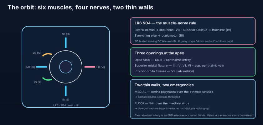
A 48-year-old woman develops a severe headache over a few hours and then notices double vision and a drooping right eyelid. When the lid is lifted, the eye is turned DOWN AND OUT — the unopposed pull of the still-working lateral rectus (VI) and superior oblique (IV) after everything the OCULOMOTOR (III) supplied has failed — and the pupil is widely DILATED and unreactive. This is a complete THIRD-NERVE PALSY, and the two features that matter most are the PAIN and the PUPIL. The parasympathetic pupil-constricting fibres run on the OUTSIDE of the oculomotor nerve, so a lesion that compresses the nerve from without — classically a POSTERIOR COMMUNICATING ARTERY ANEURYSM ballooning against it — reaches those surface fibres early and BLOWS THE PUPIL, often with pain. By contrast a medical, ischaemic palsy (from diabetes or hypertension) tends to infarct the core of the nerve and SPARE the pupil. The rule the exam wants and the on-call registrar lives by: a painful third-nerve palsy with a fixed dilated pupil is a posterior communicating aneurysm until urgent imaging proves otherwise, because the next event may be a fatal subarachnoid haemorrhage. Anatomy — which fibres sit on the surface — decides how fast the patient is scanned.
- Mixing up which nerve is which oblique. The SUPERIOR OBLIQUE is CN IV (trochlear — named for the trochlea it loops through); the INFERIOR OBLIQUE is CN III. "SO4, LR6, all the rest III" keeps it straight — the trochlear serves ONE muscle (superior oblique), the abducens ONE (lateral rectus), the oculomotor the other four plus levator.
- Treating every ptosis alike. A HEAVY ptosis with a DILATED pupil and a down-and-out eye is oculomotor (III); a PARTIAL ptosis with a CONSTRICTED pupil is sympathetic (Horner's, the superior tarsal muscle). Reading the pupil alongside the lid, not the lid alone, tells the two apart — and one of them can be an aneurysm.
- Forgetting that the ophthalmic veins have no valves. Squeezing a boil in the "danger area" of the face, or an untreated ethmoid sinusitis, can drive infection BACKWARD along valveless veins into the cavernous sinus. The orbit is not a sealed box — it drains into the skull, and infection can travel against the normal flow.
A patient has a complete right ptosis. When the lid is lifted, the eye is turned down and out and the pupil is fixed and dilated; the headache is severe. Which nerve is affected, and what does the dilated pupil most urgently suggest?
- The orbit is a four-sided bony PYRAMID with its apex at the OPTIC CANAL and base at the orbital rim, built from SEVEN bones: frontal (roof); zygomatic and greater wing of sphenoid (the strong lateral wall); maxilla, zygomatic and palatine (thin floor over the maxillary sinus); and maxilla, lacrimal, the paper-thin LAMINA PAPYRACEA of the ethmoid and sphenoid (medial wall over the ethmoid sinuses).
- Openings at the apex: OPTIC CANAL (CN II + ophthalmic artery); SUPERIOR ORBITAL FISSURE (III, IV, V1, VI + superior ophthalmic vein, sorted by the common tendinous ring); INFERIOR ORBITAL FISSURE (V2's infraorbital nerve + link to the pterygopalatine fossa); plus the ethmoidal foramina.
- Six extraocular muscles by LR6 SO4 — lateral rectus (VI), superior oblique through its trochlea (IV), the rest plus levator palpebrae (III); the smooth superior tarsal muscle is sympathetic, and its loss gives the partial ptosis of Horner's. Tears: lacrimal gland (parasympathetic from VII) → across the eye → puncta → nasolacrimal duct → inferior meatus.
- Vessels and clinical: the ophthalmic artery (from the internal carotid) gives the CENTRAL ARTERY OF THE RETINA, an end artery whose occlusion blinds; the valveless ophthalmic veins drain backward to the cavernous sinus (route of orbital/facial infection). Key scenarios: painful third-nerve palsy with a blown pupil = PCom aneurysm; blowout fracture trapping inferior rectus (up-gaze diplopia + infraorbital numbness); orbital cellulitis through the lamina papyracea; central retinal artery occlusion; cavernous sinus thrombosis; thyroid eye disease.
- Drake RL, Vogl AW, Mitchell AWM. Gray's Anatomy for Students — Head and Neck: the orbit, its walls, openings, extraocular muscles and the lacrimal apparatus.
- Moore KL, Dalley AF, Agur AMR. Clinically Oriented Anatomy — The Orbit: bony walls, superior and inferior orbital fissures, the common tendinous ring and orbital contents.
- Netter FH. Atlas of Human Anatomy — Orbit and its contents; extraocular muscles and their innervation; the lacrimal apparatus.
- Snell RS. Clinical Anatomy by Regions — The orbit: blowout fractures, the ophthalmic vessels and the cavernous sinus connections.
- Last RJ. Last's Anatomy: Regional and Applied — The orbit and eyeball; extrinsic ocular muscles and their nerve supply.
- TeachMeAnatomy — The Bony Orbit; The Extraocular Muscles; The Lacrimal Apparatus.

