
The Portal Vein: Blood That Passes Through Two Capillary Beds
Every vein you have ever drawn on a diagram does the same simple thing: it collects blood from a capillary bed and carries it home to the heart. One vein in the human body refuses. The portal vein gathers everything the gut has just absorbed — every sugar, every amino acid, every swallowed tablet, every bacterium that slipped across the wall — and instead of taking it to the heart, it delivers it to the liver, and there it breaks up into capillaries all over again. Only after the liver has read, sorted, detoxified and rationed that cargo is the blood allowed to continue. The body is checked before it is fed. That single detour is the reason a tablet swallowed is not the same drug as a tablet injected, and the reason a failing liver announces itself with swollen veins at the gullet, at the navel and at the anus — three places you would never think to look for a liver.
A man in his forties is brought into the resuscitation room having vomited what the paramedics describe, without exaggeration, as a bowl of blood. He is not in pain. That is the first strange thing — a bleed this large usually comes with an ulcer's agony, and he has none. His palms are red, there are fine spider-like vessels across his upper chest, his abdomen is tense with fluid, and a nest of dilated veins radiates from his navel like the spokes of a wheel. The registrar does not need the endoscopy report to know what has torn. Somewhere at the lower end of this man's oesophagus, a thin-walled submucosal vein — a vein that in a healthy body carries a trickle from the stomach towards the liver — has been forced to carry the pressure of an entire blocked portal system, has swollen year after year, and has finally split. The liver that should have been receiving that blood is a shrunken, scarred, unyielding block of tissue. The blood, refused entry, went looking for another way home, and the route it found runs through the wall of the gullet.
A vein that ends in capillaries
The definition of a portal system is disarmingly simple: a vein that begins in capillaries and ends in capillaries. Ordinary venous blood makes one journey through one capillary bed — tissue, vein, heart. Portal blood makes two. It leaves the capillaries of the stomach, the small and large intestine, the pancreas and the spleen; it is gathered into progressively larger veins; and then, instead of joining the inferior vena cava as every other abdominal vein does, it is delivered into the liver and broken up a second time into the hepatic sinusoids. Only when it emerges from those sinusoids into the hepatic veins does it finally reach the inferior vena cava and the heart. The consequence is that the entire nutrient harvest of a meal, and the entire absorbed dose of anything swallowed, passes under the inspection of the liver before one molecule of it reaches the brain or the heart. The liver receives about three quarters of its blood volume from the portal vein and only a quarter from the hepatic artery — but because portal blood is already partly deoxygenated, the two vessels contribute roughly equal shares of the liver's oxygen. The portal vein is not a nutrient artery to the liver; it is a delivery of goods for processing.
How the portal vein is built
Two great veins meet behind the neck of the pancreas, and a third system is born. The portal vein is formed at the level of L2, BEHIND THE NECK OF THE PANCREAS, by the union of the SPLENIC VEIN coming from the left and the SUPERIOR MESENTERIC VEIN coming up from below — a confluence that lies directly in front of the inferior vena cava and is the reason a tumour in the head or neck of the pancreas can strangle the portal circulation, as described in the duodenum and pancreas. From that confluence the vein runs upwards and to the right for about eight centimetres, entering the FREE EDGE OF THE LESSER OMENTUM — the hepatoduodenal ligament — where it occupies the deepest and most posterior position of the three great structures there: the bile duct lies in front of it and slightly to the right, the hepatic artery in front of it and slightly to the left. Behind the portal vein is the epiploic foramen, so that a finger placed in that foramen and a thumb in front of the ligament can compress all three (Pringle's manoeuvre) to stop bleeding from the liver. At the porta hepatis the vein divides into a RIGHT and a LEFT branch, the right shorter and wider, the left longer and running towards the ligamentum teres — and each branch then supplies its own half of the liver, obeying the same segmental plan as the artery and the ducts.
Its tributaries are worth naming individually, because each one becomes a clinical story later. The INFERIOR MESENTERIC VEIN, draining the hindgut, usually joins the splenic vein behind the body of the pancreas rather than the portal vein itself. The LEFT GASTRIC (coronary) vein runs along the lesser curvature and drains into the portal vein directly — it is the tributary that carries blood up to the lower oesophagus, and therefore the vein that feeds oesophageal varices. The RIGHT GASTRIC vein joins it nearby. The CYSTIC VEIN from the gallbladder drains into the right branch, and the small PARAUMBILICAL VEINS run in the falciform ligament from the umbilicus to the left branch, following the obliterated route of the fetal umbilical vein. And one negative fact governs the behaviour of the whole system: the portal vein and its tributaries have NO VALVES. There is nothing anywhere in it to stop blood running backwards. Raise the pressure at the liver and that pressure is transmitted, undamped, to every vein in the gut, the spleen and the abdominal wall connected to it.
Think of the portal vein as the customs hall of a country that imports everything it eats. Nothing arriving at the port is allowed to travel inland until it has passed through one building, been opened, inspected, taxed, repackaged or destroyed. That is the liver, and the portal vein is the single road from the docks to the customs hall. The arrangement is brilliant while the hall is working: poisons are neutralised before they reach the brain, bacteria that crossed the gut wall are eaten by resident macrophages, glucose is stored rather than flooded into the circulation. But it is also a single point of failure. Block that road — or turn the customs hall into a wall of scar tissue — and the queue does not politely wait. Goods start moving along smugglers' tracks: small unofficial paths that always existed, never carried traffic, and were never built to. Those back roads are the portosystemic anastomoses, and every dramatic sign of chronic liver disease is simply a smugglers' track overloaded.
First pass: why the route changes the drug
Because absorbed blood must cross the liver before it enters the general circulation, a drug swallowed is metabolised once before it has treated anything at all. This is FIRST-PASS METABOLISM, and for some drugs it is devastating: glyceryl trinitrate, morphine, propranolol, lidocaine and levodopa are all heavily degraded on that first pass, so the fraction of a swallowed dose reaching the arterial blood — the bioavailability — may be a small fraction of what was taken. Pharmacology answers this by choosing a route that bypasses the portal vein altogether. A tablet placed UNDER THE TONGUE drains into the lingual and facial veins, then the internal jugular and the superior vena cava — straight to the heart, no liver. A suppository placed low in the RECTUM drains through the middle and inferior rectal veins into the internal iliac veins and the inferior vena cava, escaping the portal system, whereas one that migrates high is drained by the superior rectal vein and goes to the liver after all — which is why the depth matters and why rectal bioavailability is famously erratic. Intravenous, intramuscular, transdermal and inhaled routes bypass it by definition. The enzymes doing the work are the same ones described in phase 1 and phase 2 metabolism; the anatomy simply decides whether the drug has to meet them.
The four portosystemic anastomoses
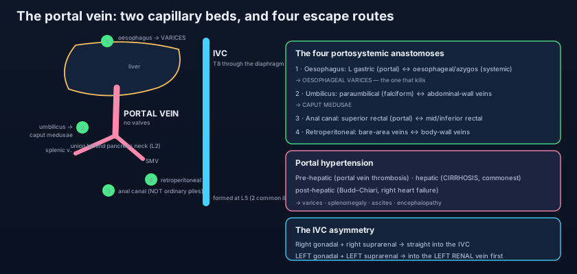
At four places in the body, a portal vein and a systemic vein touch. In health they are trivial. In portal hypertension they are everything. The FIRST and most dangerous is at the LOWER OESOPHAGUS. The oesophageal branches of the LEFT GASTRIC vein (portal) anastomose in the submucosa with the oesophageal veins draining into the AZYGOS system (systemic). When portal pressure rises, blood reverses up the left gastric vein and forces these submucosal channels open: OESOPHAGEAL VARICES. They lie immediately beneath a thin mucosa in a tube that food scrapes past several times a day, they have no muscular support, and they carry pressure they were never designed for — which is why this is the anastomosis that kills, and why it has its own management pathway in GI bleeding and oesophageal varices. The SECOND is at the UMBILICUS. The paraumbilical veins running in the falciform ligament (portal) meet the superficial epigastric, thoracoepigastric and inferior epigastric veins of the abdominal wall (systemic), described with that wall in the anterior abdominal wall. Engorged, they produce CAPUT MEDUSAE — a rosette of dilated, tortuous veins radiating from the navel, named for the snake-haired Gorgon, with blood flowing AWAY from the umbilicus in all directions.
The THIRD is at the ANAL CANAL, where the SUPERIOR RECTAL vein (portal, via the inferior mesenteric) anastomoses with the MIDDLE and INFERIOR RECTAL veins (systemic, via the internal iliac and internal pudendal). Overloaded, these produce ANORECTAL VARICES — and this deserves to be stated plainly, because it is one of the most persistent confusions in clinical medicine: anorectal varices are NOT haemorrhoids. Ordinary haemorrhoids are enlarged, prolapsing vascular cushions of the anal canal, are extremely common, and have nothing to do with portal pressure; anorectal varices are dilated portosystemic channels seen in portal hypertension, are uncommon, and are managed by lowering portal pressure rather than by banding cushions. The FOURTH is RETROPERITONEAL — the least glamorous and the most widespread. Wherever gut lies against the posterior abdominal wall without peritoneum between them, small veins of the colon and duodenum (portal) communicate with the lumbar, renal and phrenic veins of the body wall (systemic); these are the veins of Retzius, and the same principle covers the bare area of the liver, where hepatic tributaries meet phrenic veins. To these is added a fifth, occasional route: a patent DUCTUS VENOSUS, the fetal shortcut from the left portal branch to the inferior vena cava, which in some people never fully closes and can reopen under pressure.
Look for the liver at the navel. The paraumbilical veins in the falciform ligament are, embryologically, the ghost of the umbilical vein that once carried oxygenated blood from the placenta to the fetal liver. That vein closes at birth and becomes the ligamentum teres — but the tiny satellite channels beside it never entirely disappear. Decades later, when a cirrhotic liver refuses portal blood, the pressure finds them, and a route abandoned since the first minutes of life reopens in reverse. Blood that once flowed from the umbilicus inwards now flows outwards, and the veins fan out across the abdomen. In an adult with jaundice and a swollen abdomen, that rosette of veins around the navel is not a curiosity — it is an anatomical announcement that the portal system has been blocked for a long time, written on the skin.
Portal hypertension
Normal portal pressure is only about 5–10 mmHg. Very little is needed to break the system. Portal hypertension is a sustained rise in portal venous pressure, and it is classified by WHERE the obstruction sits relative to the liver. PRE-HEPATIC causes lie in the portal vein itself: portal vein thrombosis (in neonates after umbilical sepsis or catheterisation, in adults with thrombophilia, pancreatitis or malignancy) and external compression by a pancreatic tumour. HEPATIC causes lie within the liver and are by far the commonest group, dominated by CIRRHOSIS, in which regenerating nodules and fibrous septa physically distort the sinusoidal bed; schistosomiasis is a major cause worldwide. POST-HEPATIC causes lie downstream, obstructing hepatic venous outflow: Budd–Chiari syndrome (hepatic vein thrombosis), constrictive pericarditis, and right heart failure, in which a congested inferior vena cava backs pressure up into the liver. The consequences follow from a system with no valves and four escape routes. Varices open at the four anastomoses. The SPLEEN, whose only exit is the splenic vein, becomes congested and enlarges — splenomegaly, with the low platelet count of hypersplenism, as covered in the spleen. ASCITES accumulates as raised sinusoidal pressure drives fluid into the peritoneal cavity, compounded by low albumin and sodium retention. And ENCEPHALOPATHY appears because gut-derived nitrogenous waste, ammonia above all, now bypasses the liver through those same collaterals and reaches the brain unprocessed.
The man from the resuscitation room is resuscitated with blood rather than saline, started on a vasoactive drug to constrict the splanchnic arterioles and drop portal inflow, and given prophylactic antibiotics — because infection is common and worsens bleeding. Within hours he is endoscoped, and the bleeding varix is BANDED: a rubber ring is sucked over the column of vein and strangles it, so the vessel thromboses and later scars. If banding fails, a balloon tamponade tube buys a few hours. If bleeding recurs, he is offered TIPS — a transjugular intrahepatic portosystemic shunt, in which a stent is threaded from the jugular vein, down the inferior vena cava, into a hepatic vein and then pushed through the liver substance into a branch of the portal vein, creating a deliberate, controlled channel that decompresses the portal system. It works beautifully for pressure and predictably badly for the brain: the shunt does exactly what the collaterals do, so encephalopathy is its commonest complication. Long term he takes a non-selective beta-blocker to keep portal pressure down, has his ascites drained by PARACENTESIS when it splints his breathing, and joins a transplant assessment — because everything above treats the plumbing, and none of it treats the liver, a point made throughout liver disease pharmacology.
- A portal system begins in capillaries and ends in capillaries. The hepatic portal vein carries blood from the gut, pancreas and spleen to the liver sinusoids BEFORE it reaches the heart — so absorbed nutrients and drugs are processed first.
- It is formed at L2 BEHIND THE NECK OF THE PANCREAS by the SPLENIC and SUPERIOR MESENTERIC veins, is about 8 cm long, and ascends in the free edge of the lesser omentum BEHIND the bile duct and hepatic artery to divide at the porta hepatis.
- Tributaries: inferior mesenteric (usually into the splenic), left gastric/coronary (the varix-feeding vein), right gastric, cystic and paraumbilical veins. The portal vein supplies about 75% of hepatic blood flow but only half its oxygen.
- The portal system has NO VALVES — which is why a rise in pressure at the liver is transmitted backwards, unopposed, to every tributary and forces flow into collateral channels.
- The four portosystemic anastomoses: lower oesophagus (left gastric ↔ azygos) → VARICES; umbilicus (paraumbilical ↔ epigastric/thoracoepigastric) → CAPUT MEDUSAE; anal canal (superior rectal ↔ middle and inferior rectal); retroperitoneal (veins of Retzius ↔ body-wall veins), plus a patent ductus venosus.
- Portal hypertension is pre-hepatic (portal vein thrombosis), hepatic (CIRRHOSIS — commonest) or post-hepatic (Budd–Chiari, right heart failure); its consequences are varices, splenomegaly, ascites and encephalopathy.
The inferior vena cava: the vein that does go home
Set beside the portal vein, the IVC is the ordinary case — and the contrast teaches both. The inferior vena cava is formed at the level of L5 by the union of the two COMMON ILIAC VEINS, slightly to the right of the midline and just below and to the right of the aortic bifurcation described in the abdominal aorta. From there it ascends on the RIGHT SIDE of the aorta, resting on the bodies of the lumbar vertebrae and the right crus of the diaphragm, grooving the bare area on the back of the liver, and finally piercing the CENTRAL TENDON of the diaphragm at the level of T8 to enter the right atrium almost immediately afterwards — the opening detailed in the diaphragm, which cannot contract and so holds the vein permanently open. Its tributaries are read from below upwards: the common iliac veins, the median sacral vein, four pairs of LUMBAR veins, the RIGHT GONADAL vein, the two RENAL veins, the RIGHT SUPRARENAL vein, the INFERIOR PHRENIC veins and, last of all and just before it leaves the abdomen, the three HEPATIC veins. Like the portal vein it is essentially valveless, so right heart failure is transmitted straight back into the liver and the legs. And note the great negative: the inferior vena cava receives NOTHING directly from the gut. Every drop of intestinal blood must go through the liver first and can only reach the cava through the hepatic veins.
The cava is not symmetrical, and the asymmetry is entirely practical. Because the inferior vena cava lies to the RIGHT of the midline, the structures on the right reach it by a short direct route while those on the left must travel across. So the RIGHT gonadal vein (testicular or ovarian) drains directly into the cava at an acute angle, while the LEFT GONADAL VEIN drains instead into the LEFT RENAL VEIN at a right angle. In the same way the RIGHT SUPRARENAL vein is short, wide and enters the cava directly — a genuine surgical hazard during right adrenalectomy, since a torn stump retracts into the cava itself — while the LEFT SUPRARENAL vein drains into the LEFT RENAL VEIN, as set out in the adrenal glands. The left renal vein is therefore the great collector of the left side, and it must cross the midline in front of the aorta and behind the superior mesenteric artery to get there; being longer than the right renal vein, it is the reason the LEFT kidney is preferred in living donor nephrectomy — the surgeon gets more vein to sew with, a point returned to in the kidneys and ureters. Finally, when the cava itself is obstructed, blood escapes through the lumbar veins into the ASCENDING LUMBAR veins, and thence into the AZYGOS and HEMIAZYGOS system, up through the thorax and into the superior vena cava. The azygos system is the caval collateral — the systemic equivalent of the portosystemic escape routes, and the reason a slowly occluded cava can be tolerated while a sudden one cannot.
Two everyday consequences follow. The first is mechanical: because the cava is a soft, low-pressure tube lying on the vertebral bodies, anything heavy pressing on it reduces venous return. This is why a woman late in pregnancy becomes faint and pale when laid flat on her back — the gravid uterus compresses the inferior vena cava against the spine — and why she is nursed and resuscitated tilted to the LEFT. The second is oncological: because the renal veins drain straight into it, a renal cell carcinoma can grow along its own vein and put out a TUMOUR THROMBUS that climbs the inferior vena cava like a vine, sometimes as far as the right atrium, converting a kidney operation into cardiac surgery. Add to these the IVC FILTER — a small metal cage deployed percutaneously into the cava below the renal veins to catch emboli travelling up from the leg veins in a patient who cannot be anticoagulated — and the anatomy becomes something a radiologist works in daily.
- First-pass metabolism is an anatomical fact before it is a pharmacological one: swallowed drugs cross the liver before the systemic circulation, which is why glyceryl trinitrate is given sublingually and why intravenous, intramuscular, transdermal and low-rectal routes bypass the portal vein.
- The inferior vena cava is formed at L5 by the two COMMON ILIAC veins, ascends to the RIGHT of the aorta, and pierces the CENTRAL TENDON of the diaphragm at T8 — an opening that cannot contract, so it is held permanently open.
- IVC tributaries from below up: common iliac, median sacral, lumbar, right gonadal, renal, right suprarenal, inferior phrenic and hepatic veins. It has no valves and receives NOTHING directly from the gut.
- The left-sided asymmetry: the LEFT gonadal and LEFT suprarenal veins drain into the LEFT RENAL vein, not the cava; the left renal vein crosses in front of the aorta and behind the SMA, and its extra length is why the LEFT kidney is preferred in living donation.
- The right suprarenal vein is SHORT and enters the cava directly — a notorious hazard in right adrenalectomy, because a torn stump retracts into the vena cava.
- The AZYGOS system is the caval collateral: lumbar veins → ascending lumbar → azygos and hemiazygos → superior vena cava. Clinically the cava also matters in supine hypotension of pregnancy, IVC filters, and renal cell carcinoma tumour thrombus climbing towards the right atrium.
- Calling anorectal varices "haemorrhoids". Haemorrhoids are enlarged anal cushions, are very common, and are unrelated to portal pressure; anorectal varices are dilated portosystemic collaterals of portal hypertension and are treated by lowering portal pressure, not by banding cushions.
- Saying the portal vein lies in front of the bile duct in the lesser omentum. It is the most POSTERIOR of the three: bile duct in front and to the right, hepatic artery in front and to the left, portal vein behind them both.
- Assuming the gonadal and suprarenal veins are symmetrical. On the RIGHT both drain straight into the inferior vena cava; on the LEFT both drain into the left renal vein — an asymmetry that explains the left-sided varicocele and the hazards of right adrenal surgery.
A patient with cirrhosis develops dilated veins radiating from the umbilicus across the abdominal wall. Which pair of veins is anastomosing to produce this sign?
- The hepatic portal vein is formed at L2 behind the neck of the pancreas by the splenic and superior mesenteric veins, runs about 8 cm in the free edge of the lesser omentum BEHIND the bile duct and hepatic artery, and divides at the porta hepatis — carrying gut, pancreatic and splenic blood through a SECOND capillary bed in the liver before it ever reaches the heart.
- It has no valves, so portal hypertension — pre-hepatic (portal vein thrombosis), hepatic (cirrhosis, commonest) or post-hepatic (Budd–Chiari, right heart failure) — is transmitted backwards and forces blood into four portosystemic anastomoses, producing varices, splenomegaly, ascites and encephalopathy.
- The four anastomoses: lower oesophagus (left gastric ↔ azygos) giving the varices that kill; umbilicus (paraumbilical ↔ epigastric/thoracoepigastric) giving caput medusae; anal canal (superior rectal ↔ middle and inferior rectal) giving anorectal varices, which are NOT haemorrhoids; and retroperitoneal (veins of Retzius ↔ body-wall veins).
- The inferior vena cava is the ordinary vein by contrast: formed at L5 from the two common iliac veins, ascending to the RIGHT of the aorta and piercing the diaphragm's central tendon at T8; valveless, receiving nothing directly from the gut, and asymmetric — the LEFT gonadal and LEFT suprarenal veins drain into the LEFT RENAL vein, which is why the left kidney is preferred for donation.
- Drake RL, Vogl AW, Mitchell AWM. Gray's Anatomy for Students — Abdomen: the hepatic portal system and portosystemic anastomoses.
- Moore KL, Dalley AF, Agur AMR. Clinically Oriented Anatomy — Portal vein, portal hypertension and the inferior vena cava.
- Netter FH. Atlas of Human Anatomy — Venous drainage of the abdomen; portal vein tributaries and portocaval anastomoses.
- Last RJ. Last's Anatomy: Regional and Applied — The portal vein and the posterior abdominal wall veins.
- Snell RS. Clinical Anatomy by Regions — Oesophageal varices, caput medusae and inferior vena caval obstruction.
- TeachMeAnatomy — The Hepatic Portal System; The Inferior Vena Cava.

