
The Liver: The Only Organ That Grows Back
Cut away most of a lung and it will never come back. Cut away most of a kidney, a pancreas, a piece of brain — gone for good, replaced at best by scar. Now take a healthy adult, remove two thirds of the liver, and within a few weeks the remaining third has grown until the organ is very nearly the size it was before. That single fact is why a living person can donate a lobe of their liver to a dying stranger and both of them walk out of hospital with a whole liver each. It is the largest internal organ, about a kilogram and a half, it takes a quarter of everything the heart pumps, and it is plumbed in a way no other organ is: two blood supplies coming in, three veins going out, and a secret internal geography that has nothing to do with the lumps you can see from the outside.
Two operating tables, two teams, one morning. On the first table a man in his fifties is asleep with his abdomen open; his own liver is a hard, shrunken, nodular thing that no longer works, and the veins on his belly wall stand out like cords. On the second table lies his daughter, thirty-one years old and entirely healthy, who has volunteered to give him the right side of her liver. The surgeon operating on her does not cut along the obvious landmark — the pale falciform ligament running down the front of the organ like a seam. She cuts along a line no eye can see, drawn in the mind from the notch of the gallbladder bed to the inferior vena cava, and follows the middle hepatic vein down through the parenchyma. Bleeding is controlled by a soft clamp on a single bundle in the free edge of the lesser omentum. By evening the daughter has half a liver and the father has the other half, and each of those halves will grow. It works because the liver's true anatomy is not the anatomy of its surface.
Where it sits, and what it touches
You cannot normally feel a healthy liver, because the ribs are hiding it. The liver fills the right hypochondrium, spills across the epigastrium and reaches a short distance into the left hypochondrium, moulded from above into the domes of the diaphragm and shielded almost entirely by ribs 7 to 11 — which is why a normal liver edge is not palpable below the costal margin, and why a liver you can easily feel is either enlarged or pushed down. It has two surfaces. The DIAPHRAGMATIC surface is smooth and convex, following the curve of the diaphragm over the front, above and behind. The VISCERAL surface faces down and backwards and is anything but smooth: it is dented by everything it lies on — the gallbladder, the right kidney and right adrenal gland, the duodenum, the right colic flexure, the stomach and the oesophagus each leave a named impression in it. Between the two, the sharp INFERIOR BORDER runs from right to left across the abdomen; it is this edge that a clinician tries to catch with the fingertips at the height of a deep breath, because the liver descends with every inspiration.
One patch of the diaphragmatic surface is different, and it matters more than its size suggests. The liver is almost completely wrapped in visceral peritoneum, but on its posterosuperior aspect there is a triangular region where the peritoneum is reflected away onto the diaphragm and the liver capsule lies directly against diaphragmatic muscle: the BARE AREA. Its boundary is the CORONARY LIGAMENT — simply the line along which the peritoneum turns back — whose right and left extremities come together as the right and left triangular ligaments. Because there is no peritoneal layer here, the bare area is a direct anatomical bridge between liver and diaphragm, and infection or collection tracking along it reaches the subphrenic space above. It also explains why a hepatic abscess can irritate the diaphragm and be felt at the tip of the right shoulder, and why the inferior vena cava, buried in a deep groove in the back of the bare area, is in effect inside the liver rather than merely behind it.
The anatomical lobes — an old and misleading map
The divisions you can see with your eyes are the ones that matter least. Looked at from the front, the liver appears to be split into a huge RIGHT LOBE and a much smaller LEFT LOBE by the falciform ligament, a thin peritoneal fold running from the anterior abdominal wall to the diaphragmatic surface and carrying the LIGAMENTUM TERES — the obliterated left umbilical vein — in its free lower edge. Turn the organ over and the visceral surface shows two more lobes, laid out in a pattern generations of students have memorised as the letter H. The left limb of the H is the fissure for the ligamentum teres in front and the fissure for the LIGAMENTUM VENOSUM (the obliterated ductus venosus) behind. The right limb is the gallbladder fossa in front and the groove for the INFERIOR VENA CAVA behind. The crossbar joining them is the PORTA HEPATIS, the doorway through which the vessels and ducts enter and leave. The block of liver in front of the crossbar, between gallbladder and ligamentum teres, is the QUADRATE LOBE; the block behind it, between the inferior vena cava and the ligamentum venosum, is the CAUDATE LOBE. It is a tidy description of a surface — and as a guide to how the liver actually works, it is close to useless.
Cantlie's line: the liver's real midline
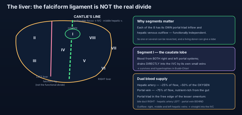
Ask not what the surface shows, but where the blood divides. The functional right and left livers are defined by their blood, not by their bumps. The portal vein, the hepatic artery and the bile duct each divide into a right and a left branch at the porta hepatis, and the territory each set of branches supplies is a true half — a hemiliver. The plane between those two halves is CANTLIE'S LINE: an imaginary sagittal plane running from the middle of the gallbladder fossa in front, backwards to the left side of the inferior vena cava behind, with the MIDDLE HEPATIC VEIN lying within it as the internal marker. Notice what this means. Cantlie's line passes well to the RIGHT of the falciform ligament, so the quadrate lobe — which looks as though it belongs to the right lobe — is functionally part of the LEFT liver, and most of the caudate lobe is functionally separate from both. The surgeon who wants to take the right half of a liver therefore ignores the falciform ligament entirely and transects along Cantlie's plane, because that is the only line along which the two halves can be separated without robbing either of its blood supply.
Eight segments, eight little livers
Cantlie's line is only the first cut. Follow the branches further and the liver resolves into EIGHT COUINAUD SEGMENTS, each of which receives its own portal triad — a branch of the portal vein, a branch of the hepatic artery and a tributary bile duct — and each of which is drained at its margins by the hepatic veins. The two great vertical planes are the three hepatic veins (right, middle and left), and the horizontal plane is the level of the right and left portal branches, dividing each sector into a superior and an inferior segment. Numbered clockwise as the liver is seen from the front, segment I is the caudate lobe, segments II and III form the lateral part of the left liver, segment IV the medial part (the old quadrate lobe, often split into IVa above and IVb below), and segments V to VIII make up the right liver — V and VIII in the anterior sector, VI and VII in the posterior. The consequence is the single most useful idea in liver surgery: because each segment has its own inflow and its own outflow, one or several can be removed and the remainder keeps its blood, its bile drainage and its function. A tumour confined to segment VI can be taken out and the other seven segments never know it happened.
Think of the liver not as one organ but as a block of eight flats, built around a central stairwell. The stairwell is the portal triad rising from the porta hepatis; each flat is served by its own branch off it — its own water main, its own electricity, its own waste pipe — and empties its used water into drains buried in the party walls, which are the hepatic veins. Demolish one flat and the others carry on, because nothing essential to them passed through it. This is exactly why liver resection is possible and, say, resection of a piece of the pancreas is so much harder: the neighbours in the liver are independent tenants, not roommates sharing a single supply. And the one flat that breaks the rule is the ground-floor apartment, segment I — it has its own private connections to both stairwells and its own private drain straight out to the street.
The caudate lobe is the liver's escape hatch. Segment I is the only segment that receives portal blood from BOTH the right and the left portal systems, and — uniquely — it does not drain into the three main hepatic veins at all. Instead it empties by its own short veins directly into the inferior vena cava lying immediately behind it. Now consider Budd–Chiari syndrome, in which the hepatic veins thrombose and the liver strangles on its own outflow: every other segment congests, swells and dies, while the caudate, draining by a private route the clot never reached, survives — and then hypertrophies, sometimes enormously, as it takes on the work of the rest. A grossly enlarged caudate lobe on a scan of a congested liver is not a curiosity; it is close to a diagnosis.
Two supplies in, three veins out
No other organ in the body is fed by two entirely different rivers. The HEPATIC ARTERY PROPER carries oxygenated blood: it is the continuation of the common hepatic artery, itself one of the three branches of the coeliac trunk described in the abdominal aorta, after the gastroduodenal artery has been given off. It supplies only about a quarter of the liver's blood flow, but because that blood is fully oxygenated it delivers roughly half of the liver's oxygen — and it is the sole supply of the bile ducts, which is why hepatic artery thrombosis after a transplant destroys the biliary tree first. The PORTAL VEIN carries the other three quarters: dark, deoxygenated, but loaded with everything absorbed from the gut, formed behind the neck of the pancreas by the union of the splenic and superior mesenteric veins, as traced in the portal system and inferior vena cava. Every molecule of food, drug or toxin absorbed from the intestine is presented to the hepatocytes before it is allowed anywhere near the systemic circulation — the anatomical basis of first-pass metabolism and of everything in phase 1 and phase 2 metabolism.
The two vessels and the bile duct do not travel alone. They run together — as they will continue to do all the way down to the smallest branch inside the liver — in the free edge of the lesser omentum, the double fold of peritoneum described in the mesenteries and omenta, where they form the PORTAL TRIAD. The arrangement at the free edge is worth learning as three words: DUCT right, ARTERY left, VEIN behind. Behind the triad lies the omental (epiploic) foramen of Winslow, the only natural doorway into the lesser sac, and a surgeon can slip a finger through it, pinch the free edge between finger and thumb and arrest almost all blood flow into the liver at once. That is the PRINGLE MANOEUVRE, described in 1908 and still performed today: it stops bleeding from the hepatic artery and the portal vein together, and — crucially — bleeding that continues despite a Pringle clamp is coming from the hepatic veins or the inferior vena cava behind, not from the inflow.
Outflow is simpler and far more dangerous. Blood leaves the liver through THREE HEPATIC VEINS — right, middle and left — which run inside the parenchyma in the planes BETWEEN the segments (the intersegmental planes, in contrast to the portal triads which run within the segments) and empty into the inferior vena cava at the top of the bare area, just below the diaphragm; the left and middle commonly join as a short common trunk before they enter. The critical practical point is that there is essentially no extrahepatic hepatic vein: the veins are only a centimetre or so long outside the liver substance before they disappear into the cava, so there is nothing to clamp. A tear here bleeds into the inferior vena cava under negative intrathoracic pressure, and the same anatomy that makes the liver easy to divide makes its outflow one of the most feared injuries in trauma surgery. Add the small independent veins of the caudate lobe going straight into the cava, and you have the complete picture: two rivers in through one gate, three rivers out through the back wall.
- The liver occupies the right hypochondrium and epigastrium under the diaphragm, shielded by ribs 7–11; it has a smooth diaphragmatic surface and an indented visceral surface, and moves down with every inspiration.
- The BARE AREA on the posterosuperior diaphragmatic surface has no peritoneum and is bounded by the coronary ligament — a direct route from liver to the subphrenic space.
- The ANATOMICAL lobes (right, left, caudate, quadrate) are surface divisions made by the falciform ligament and the H-shaped grooves: ligamentum teres and ligamentum venosum on the left limb, gallbladder fossa and IVC groove on the right, porta hepatis as the crossbar.
- CANTLIE'S LINE — gallbladder fossa to inferior vena cava, along the middle hepatic vein — is the true division into functional right and left livers; the quadrate lobe therefore belongs to the LEFT liver.
- EIGHT COUINAUD SEGMENTS, numbered clockwise from the front with segment I the caudate: each has its own portal triad inflow and hepatic venous outflow, so any one can be resected independently.
- Segment I (caudate) is unique: portal blood from BOTH sides and its own small veins draining straight into the IVC — hence it survives and hypertrophies in Budd–Chiari syndrome.
Inside a segment: the lobule and the acinus
Zoom in far enough and the same two-rivers-in, one-river-out pattern repeats. The classic histological unit is the HEPATIC LOBULE: a roughly hexagonal column of hepatocytes arranged in plates radiating around a CENTRAL VEIN, with a portal triad — bile duct, hepatic arteriole and portal venule — at each of the six corners. Blood runs from the corners towards the centre through SINUSOIDS, wide capillaries lined by an unusual FENESTRATED endothelium with no basement membrane, so that plasma bathes the hepatocytes almost directly across the perisinusoidal SPACE OF DISSE, where the stellate (Ito) cells that store vitamin A — and that turn into the collagen-laying myofibroblasts of cirrhosis — reside. Fixed macrophages, the KUPFFER CELLS, sit within the sinusoidal lining and strip bacteria and old red cells out of portal blood before it reaches the systemic circulation. Bile, meanwhile, travels the opposite way: hepatocytes secrete it into tiny canaliculi between their facing surfaces, and it flows outwards, against the direction of the blood, towards the bile ductules at the corners and on into the system described in the biliary tree and gallbladder.
The lobule is a beautiful drawing, but the functionally honest unit is the PORTAL ACINUS: a diamond of tissue centred not on the central vein but on the portal triad, divided into three zones by distance from the incoming blood. ZONE 1, periportal, meets the blood first — best oxygenated, richest in the enzymes of oxidative metabolism, and therefore the first tissue struck by an ingested toxin and the site of most viral and biliary injury. ZONE 3, centrilobular, sits farthest away next to the central vein — the last to be oxygenated and the first to die when perfusion falls, which is why shock, heart failure and hypotension produce centrilobular necrosis and the mottled "nutmeg liver" of chronic congestion. Zone 3 is also where cytochrome P450 activity is concentrated, so a drug that is toxic only after it has been metabolised destroys zone 3 selectively: this is exactly the pattern of paracetamol necrosis in overdose, where the reactive metabolite NAPQI overwhelms glutathione precisely in the zone that made it.
Held in place by folds of peritoneum
The liver is an intraperitoneal organ, and its attachments are all condensations of the membrane described in the peritoneum. In front, the FALCIFORM LIGAMENT tethers it to the anterior abdominal wall and the underside of the diaphragm, carrying the ligamentum teres in its free edge — a remnant that becomes clinically loud in portal hypertension, when the paraumbilical veins running alongside it reopen and produce caput medusae. Above and behind, the two layers of the falciform ligament separate to become the CORONARY LIGAMENT, enclosing the bare area, and where its layers meet at the right and left extremes they form the TRIANGULAR LIGAMENTS — the left one long and easily divided, the right short and tucked deep. Below, the LESSER OMENTUM runs from the porta hepatis and the fissure for the ligamentum venosum to the lesser curvature of the stomach and the first part of the duodenum, so its two named parts are the hepatogastric and hepatoduodenal ligaments; the hepatoduodenal ligament is the free edge, and the free edge is the portal triad. Note the elegance of the arrangement: the same fold that anchors the liver to the stomach also delivers its blood and takes away its bile.
The examining hand: a doctor places the right hand flat below the costal margin, presses gently, and asks the patient to breathe in; the descending liver edge bumps against the fingertips. She measures the span by percussion, notes whether the edge is soft or hard, smooth or knobbly, tender or painless, pulsatile or still. The donating hand: a living-donor hepatectomy takes segments V–VIII along Cantlie's plane, leaving the donor with segments I–IV — the left liver — which will regenerate to nearly full volume within weeks. The biopsying hand: a needle is passed in the mid-axillary line through the eighth or ninth intercostal space at the end of EXPIRATION, above the costal margin but below the pleural reflection, because the liver rides up in expiration and the pleura dips down in inspiration. The clamping hand: a trauma surgeon whose patient is exsanguinating from a shattered liver puts a finger through the epiploic foramen and performs a Pringle manoeuvre — and if the bleeding does not stop, knows instantly that the injury is in the hepatic veins behind.
When the architecture is destroyed
Cirrhosis is not a disease of hepatocytes so much as a disease of ARCHITECTURE. Repeated injury — alcohol, viral hepatitis, fatty liver — activates the stellate cells of the space of Disse, which lay down collagen, the sinusoids lose their fenestrations, and the parenchyma is remodelled into regenerative nodules strangled by bands of fibrous septa. Portal blood can no longer flow freely through the sinusoids to the central veins, and pressure in the portal vein rises. Since that vein has no valves, blood escapes backwards through portosystemic anastomoses: at the lower oesophagus (varices, the subject of GI bleeding and oesophageal varices), at the umbilicus (caput medusae, along the reopened paraumbilical veins of the ligamentum teres), at the rectum, and in the retroperitoneum. The liver itself may shrink while the spleen enlarges and ascites collects. Meanwhile the loss of hepatocyte mass ruins synthesis and detoxification, and the failing organ becomes a pharmacological problem in its own right, as set out in liver disease pharmacology.
The liver has almost no pain fibres in its substance — you can burn, cut and biopsy hepatic parenchyma and the patient feels nothing. The pain of liver disease comes from GLISSON'S CAPSULE, the fibrous coat around it, which is innervated by the lower intercostal nerves and stretches when the organ swells rapidly. That single fact explains a whole clinical pattern: acute hepatitis, right heart failure engorging the liver overnight, or a fast-growing abscess all cause a dull ache and tenderness in the right hypochondrium, whereas cirrhosis — which develops over years and may even shrink the liver — is famously painless until it decompensates. A liver that hurts is a liver that grew fast. And where the swollen liver touches the diaphragm over the bare area, that pain may be referred to the tip of the right shoulder along C3–C5 instead.
- DUAL supply: hepatic artery proper (~25% of flow, ~50% of oxygen, sole supply of the bile ducts) and portal vein (~75% of flow, nutrient-rich from the gut).
- PORTAL TRIAD in the free edge of the lesser omentum: bile DUCT right, ARTERY left, portal VEIN behind — pinched in the Pringle manoeuvre through the epiploic foramen.
- Three hepatic veins (right, middle, left) run BETWEEN segments and enter the IVC with essentially no extrahepatic length — nothing to clamp, so outflow bleeding is dangerous.
- Microstructure: hexagonal lobule with a central vein and six corner triads; fenestrated sinusoids, Kupffer cells, and the space of Disse with its stellate cells (the source of cirrhotic collagen).
- Portal acinus zones: ZONE 1 periportal is best oxygenated and hit first by ingested toxins; ZONE 3 centrilobular is least oxygenated — the site of ischaemic necrosis and of paracetamol (NAPQI) necrosis.
- Peritoneal attachments: falciform (with ligamentum teres), coronary (bounding the bare area), the two triangular ligaments, and the lesser omentum (hepatogastric + hepatoduodenal).
- Believing the falciform ligament divides the liver into right and left halves. It divides the SURFACE only; the functional division is Cantlie's line from the gallbladder fossa to the IVC — which is why the quadrate lobe belongs to the left liver, not the right.
- Assuming the portal vein delivers most of the liver's oxygen because it delivers most of its blood. It carries about 75% of the flow but only about half the oxygen; lose the hepatic artery and the bile ducts, which have no other supply, necrose first.
- Expecting hepatic veins to follow the portal triads. The triads run WITHIN segments; the hepatic veins run BETWEEN them, in the intersegmental planes — that opposition is the entire basis of segmental resection.
A surgeon planning a right hepatectomy transects the liver along a plane running from the middle of the gallbladder fossa backwards to the inferior vena cava, rather than along the falciform ligament. Which structure lies within that plane and defines it?
- The liver (~1.5 kg, a quarter of the cardiac output) fills the right hypochondrium and epigastrium under the diaphragm behind ribs 7–11, with a diaphragmatic and a visceral surface and a peritoneum-free BARE AREA bounded by the coronary ligament that opens onto the subphrenic space.
- The anatomical lobes (right, left, quadrate, caudate) drawn by the falciform ligament and the H-shaped grooves are a surface myth; the TRUE division is Cantlie's line — gallbladder fossa to IVC along the middle hepatic vein — and the EIGHT COUINAUD SEGMENTS, each with its own portal inflow and hepatic venous outflow, which is what makes segmental resection possible.
- Segment I (caudate) takes portal blood from both sides and drains directly into the IVC, so it survives and hypertrophies in Budd–Chiari; inflow is the hepatic artery proper (25% flow, 50% oxygen) plus the portal vein (75%) travelling as the portal triad — duct right, artery left, vein behind — clamped in the Pringle manoeuvre; outflow is three hepatic veins with no extrahepatic length.
- Microscopically the hexagonal lobule drains to a central vein through fenestrated sinusoids with Kupffer cells and the space of Disse, while the portal acinus grades zone 1 (periportal, best oxygenated, first hit by ingested toxins) to zone 3 (centrilobular, the site of ischaemic and paracetamol necrosis) — and clinically the liver is painless except when Glisson's capsule is stretched, regenerates after resection, and fails architecturally in cirrhosis with portal hypertension.
- Drake RL, Vogl AW, Mitchell AWM. Gray's Anatomy for Students — Abdomen: the liver, its surfaces, lobes and segments.
- Moore KL, Dalley AF, Agur AMR. Clinically Oriented Anatomy — The liver: peritoneal relations, portal triad, hepatic segmentation and the Pringle manoeuvre.
- Netter FH. Atlas of Human Anatomy — Liver: surfaces, vasculature and segmental anatomy.
- Last RJ. Last's Anatomy: Regional and Applied — The liver and the extrahepatic biliary apparatus.
- Snell RS. Clinical Anatomy by Regions — Hepatomegaly, liver biopsy, subphrenic spaces and portal hypertension.
- Couinaud C. Le Foie: Études Anatomiques et Chirurgicales — the segmental anatomy of the liver.
- TeachMeAnatomy — The Liver; The Portal Venous System.

