
The Heart: Two Pumps in One Fist
Close your hand and look at it. That is roughly the size of your heart — a hollow muscle no bigger than your own fist, sitting behind the breastbone, that has been contracting since the fourth week of your life in the womb and will not stop until the last minute of it. About 100,000 beats a day. Some 2.5 billion in a lifetime. No holiday, no night shift handover, no rest longer than the fraction of a second between one beat and the next. And the strangest thing about it is that it is not one pump at all. It is two pumps, bolted together, sharing a common wall, working in perfect step — one sending blood a few centimetres to the lungs, the other driving it to the tip of your toe.
A newborn is placed on her mother's chest, and in the first few breaths something reorganizes inside her that will never be undone. For nine months a hole in the wall between her two atria — the foramen ovale — let blood bypass lungs that were full of fluid and did no breathing. Now the lungs inflate, pulmonary resistance collapses, pressure in the left atrium rises above the right, and a flap of tissue is pressed shut against the septum like a door closing in a draught. Within months it will seal, leaving only a shallow dimple that anatomists call the fossa ovalis. Put your hand on that child's chest sixty years later, in the fifth intercostal space just inside the nipple line, and you can feel the tap of her apex beat: the tip of a left ventricle whose wall is now three times thicker than the right, still pushing against a door that closed on the day she was born.
How the heart actually sits in the chest
Almost every drawing of the heart lies about its position. The heart is drawn upright and central; in reality it lies obliquely, tipped forward and to the left, with about two-thirds of its mass to the LEFT of the midline. It occupies the middle mediastinum, wrapped in its own serous sac — the subject of the pericardium — inside the middle compartment of the mediastinum. Orient yourself by two poles. The APEX points down, forward and to the left, and lies in the fifth intercostal space in the mid-clavicular line; it is formed entirely by the LEFT VENTRICLE, and it is what you feel as the apex beat. The BASE faces backwards, upwards and to the right, and is formed mostly by the LEFT ATRIUM (with a small contribution from the right atrium) — which is why the left atrium, the chamber you can least easily picture, is the one lying directly against the oesophagus and the vertebral column.
Between those two poles the heart presents surfaces and borders, and each one is dominated by a different chamber — a fact that turns a plain chest radiograph into a readable map. The anterior (sternocostal) surface is mostly RIGHT VENTRICLE: the chamber closest to your breastbone is not the left ventricle everyone imagines, but the right, which is why a sternal blow or a needle passed under the xiphoid reaches the right ventricle first. The inferior (diaphragmatic) surface, lying on the central tendon of the diaphragm, is formed by both ventricles, mainly the left. The left (pulmonary) surface, filling the cardiac notch of the left lung, is left ventricle. The right border is RIGHT ATRIUM; the left border is LEFT VENTRICLE with the left auricle; the inferior border is chiefly right ventricle; and the superior border is the two atria and their auricles, with the great vessels leaving between them.
The grooves on the outside — and the crux
Look at a heart from outside and you see grooves, filled with fat, that mark exactly where the internal walls lie — and, conveniently, carry the vessels described in the coronary arteries and cardiac veins. The coronary (atrioventricular) sulcus runs around the heart like a slanted belt, separating the atria above from the ventricles below; it lodges the right coronary artery, the circumflex artery and the coronary sinus. The anterior interventricular sulcus runs down the front from that belt to a notch just right of the apex, marking the interventricular septum and carrying the anterior interventricular (LAD) artery. The posterior interventricular sulcus does the same on the diaphragmatic surface. Where the coronary sulcus, the posterior interventricular sulcus and the interatrial groove all converge on the back of the heart is the CRUX — the anatomical crossroads where all four chambers meet, and the point from which the artery to the atrioventricular node usually arises. A blocked vessel at the crux therefore threatens not just muscle but the wiring.
The wall itself is three layers, and each one has a disease named after it. From inside out: the ENDOCARDIUM, a smooth endothelial lining continuous with the endothelium of the great vessels, which also covers the valve cusps — inflame or infect it and you have endocarditis, with vegetations growing on those cusps. The MYOCARDIUM, the thick middle layer of cardiac muscle arranged in spiralling sheets that wring the ventricles rather than merely squeezing them; this is the layer that dies in a myocardial infarction and hypertrophies in hypertension. And the EPICARDIUM, which is not a separate coat at all but the visceral layer of serous pericardium applied directly to the heart, with the coronary vessels and their fat running just beneath it. Understanding that the epicardium IS the visceral pericardium is what makes the pericardial cavity, and the tamponade that can fill it, make sense.
The right atrium: a chamber of two halves
Open the right atrium and you find, unexpectedly, two completely different interiors welded together — a fossil record of how it was built. The posterior part is the SINUS VENARUM: perfectly smooth-walled, developed from the embryonic sinus venosus, and it is the collecting basin. Into it drain the superior vena cava (from above, no valve), the inferior vena cava (from below, guarded by the rudimentary valve of the IVC — the Eustachian valve, which in fetal life directed oxygenated placental blood across the foramen ovale) and the coronary sinus, returning the heart's own venous blood between the IVC orifice and the tricuspid opening. The anterior part, including the ear-shaped right AURICLE, is rough: ridged by parallel bundles of PECTINATE MUSCLES like the teeth of a comb. Between the two runs a vertical muscular ridge, the CRISTA TERMINALIS, matched on the outside by a shallow groove, the sulcus terminalis. That crest is not merely a landmark — at its upper end, near the mouth of the superior vena cava, sits the sinu-atrial node, the pacemaker described in the conducting system of the heart.
On the septal wall of the right atrium is the FOSSA OVALIS, an oval depression with a prominent upper and anterior rim, the limbus. It is the scar of the foramen ovale — the fetal shunt whose closure you watched in the opening scene — and it is the thinnest part of the interatrial septum, which is why cardiologists cross it deliberately to reach the left side of the heart. In roughly a quarter of adults the flap never fully fuses, leaving a probe-patent foramen ovale. Most of the time this means nothing at all. Occasionally it means a great deal: a clot formed in a leg vein, which should end its journey harmlessly in the lung, can be pushed through the flap when right atrial pressure transiently exceeds left — a strain, a cough, a Valsalva — and travel instead to the brain. That is paradoxical embolism, a venous clot causing an arterial stroke, and its entire mechanism is one unclosed door in this wall. Lower on the septal wall, near the coronary sinus orifice, lies the small triangle of Koch, which contains the atrioventricular node.
The right ventricle: the low-pressure pump
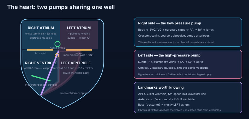
It only has to push blood as far as the lungs — and its whole design says so. In cross-section the right ventricle is a crescent, wrapped around the bulging left ventricle rather than being a chamber in its own right, and its wall is only about 3 to 5 mm thick. That thinness is not weakness; it is economy. Pulmonary vascular resistance is a fraction of systemic resistance, so this pump generates a systolic pressure of roughly 25 mmHg where the left generates 120. Inside, the inflow portion is heavily ridged by coarse TRABECULAE CARNEAE, and three PAPILLARY MUSCLES — anterior (the largest), posterior and a small septal group — rise from the wall. From their tips, fine tendinous cords, the CHORDAE TENDINEAE, fan out to the free edges of the tricuspid cusps. Their job is not to open or close the valve but to tether it: when the ventricle contracts, the papillary muscles contract with it and hold the cusps down so that they cannot prolapse back into the atrium, exactly as detailed in heart valves and heart sounds.
Two structures make the right ventricle instantly recognizable. The first is the MODERATOR BAND, properly the septomarginal trabecula: a stout muscular bridge that leaps across the cavity from the interventricular septum to the base of the anterior papillary muscle. Its old name came from the belief that it moderated over-distension of the chamber, but its real importance is electrical — it carries the right bundle branch across the cavity so that the anterior papillary muscle is excited early and takes up tension before the rest of the ventricle contracts. On an echocardiogram it is the reliable marker that says "this chamber is the right ventricle." The second is the CONUS ARTERIOSUS, or infundibulum: a smooth, funnel-shaped outflow tract in the upper part of the chamber, separated from the rough inflow by the supraventricular crest and leading up to the pulmonary valve. Rough inflow, smooth outflow — a pattern you will meet again on the left.
Think of the two sides of the heart as two pumps on the same shaft in a building's water system. The right-hand pump only has to lift water to the floor above — a short, low-pressure job — so it is built light: a thin crescent-shaped casing, cheap to run, and it would burst if you asked it to do more. The left-hand pump has to drive water to the roof of a tower block and out to every tap in the building, so it is a heavy conical cylinder with walls two to three times thicker. Both must move exactly the same volume every stroke, or the building floods: pump a little more on the right for long enough and fluid backs up into the lungs. The wall between them is not a partition added afterwards but a shared structural member — which is why a hole in it, a ventricular septal defect, does not merely leak; it lets the strong pump drive its output straight into the weak one.
The left atrium and the left ventricle
The LEFT ATRIUM is the quietest chamber and the hardest to see: it forms most of the base of the heart, lying against the oesophagus, which is exactly why a transoesophageal echo probe gives such superb pictures of it. Its walls are almost entirely smooth, because it develops largely by absorbing the primitive pulmonary vein, and into its posterior wall open the FOUR PULMONARY VEINS — two right, two left — without any valves. Only its small ear-like appendage, the left auricle, retains pectinate muscles, and that little trabeculated pouch matters out of all proportion to its size: in atrial fibrillation, when the atrium quivers instead of contracting, blood stagnates in the appendage, and it becomes the commonest site of thrombus formation in the whole heart. A clot forming there sits in the systemic circuit; when it leaves, it travels to the brain. Anticoagulation in AF, and the rate-versus-rhythm decisions covered in atrial fibrillation and SVT, are ultimately about this one small pouch.
Everything about the left ventricle is an answer to one question: how do you generate pressure? The LEFT VENTRICLE is conical, its cavity circular in cross-section, and its wall is 8 to 12 mm thick — two to three times the right — because it must drive blood through the entire systemic circuit, from the aortic root to the capillaries of your toes and back. Its trabeculae carneae are finer and more numerous than the right ventricle's, and it has only TWO papillary muscles, both large: the anterolateral and the posteromedial. That asymmetry has a price. The anterolateral papillary muscle usually enjoys a dual blood supply (from the LAD and the circumflex), while the posteromedial is typically supplied by a single vessel — so in an inferior myocardial infarction it is the posteromedial muscle that ruptures, tearing the mitral apparatus and producing sudden, catastrophic mitral regurgitation days after the infarct. Above the anterior mitral cusp the wall becomes smooth again: the aortic vestibule, the fibrous outflow tract leading to the aortic valve and the aortic root.
The septa and the fibrous skeleton
Two septa divide the heart. The INTERATRIAL SEPTUM is thin and largely muscular, thinning further at the fossa ovalis. The INTERVENTRICULAR SEPTUM is a different beast: overwhelmingly a thick MUSCULAR part, functionally belonging to the left ventricle and bulging into the right, plus a small MEMBRANOUS part high up and posteriorly, where it meets the fibrous skeleton. That membranous portion is only a sheet of fibrous tissue a few millimetres across, and it does two disproportionately important things. It is the commonest site of a ventricular septal defect — the commonest congenital heart defect of all — because it is the last part of the septum to close in development, formed by the fusion of three separate contributions. And it is the doorway through which the atrioventricular bundle of His passes from atrium to ventricle. A perimembranous VSD, or surgery to repair one, therefore risks heart block, because the hole and the wire share a postcode.
Holding all of this together is a structure students routinely skip and examiners routinely love: the FIBROUS SKELETON of the heart. It is a set of four dense fibrous rings — the annuli fibrosi — encircling the two atrioventricular orifices and the aortic and pulmonary orifices, joined by the right and left fibrous trigones and continuous with the membranous septum. It does three things at once. It gives the valve cusps and the myocardium of both atria and ventricles something to attach to, so the muscle has an anchor rather than pulling on other muscle. It keeps the valve orifices from stretching open as the chambers dilate. And, most elegantly, it is electrically inert — a complete insulating gasket between atrial and ventricular muscle, so that a wave of depolarization sweeping through the atria cannot cross directly into the ventricles. The only permitted route is the atrioventricular bundle, piercing the skeleton at one point. Without that insulation, the atrioventricular delay that lets the atria finish filling the ventricles could not exist, and a fast atrial rhythm would drive the ventricles beat for beat.
The single most useful number in cardiac anatomy is the ratio 3:1. The left ventricular wall is roughly three times the thickness of the right, and it is not because the left ventricle moves more blood — the two ventricles must eject identical volumes, or the circulation fails within minutes. It is because of pressure. Muscle thickness in the heart is a record of the pressure a chamber has spent its life working against, which is why the whole thing runs in reverse in disease: give the left ventricle years of untreated hypertension and it thickens further still (concentric hypertrophy, and the apex beat becomes forceful and sustained); give the right ventricle years of pulmonary hypertension or a chronic lung disease and the thin crescent thickens into something that begins to resemble the left — cor pulmonale. Read a heart's walls and you are reading its biography.
Feel your own apex beat: lie on your left side, find the fifth intercostal space in the mid-clavicular line, and the light tap under your fingers is the left ventricle. If it is displaced down and out towards the axilla, the ventricle is dilated; if it is a forceful, sustained heave in the normal place, it is hypertrophied — the fingertip diagnosis of long-standing hypertension, whose muscle cost eventually shows up as the failing heart treated in heart failure: the four pillars. A 74-year-old with an irregularly irregular pulse has atrial fibrillation; the stroke he is at risk of will most likely begin as a clot in a left atrial appendage that has not truly contracted for years. A young woman with a stroke and no vascular risk factors at all gets a bubble study, and bubbles injected into an arm vein appear in her left atrium within three beats — a patent foramen ovale, and paradoxical embolism. And a baby with a loud harsh pansystolic murmur at the left sternal edge and poor feeding has a perimembranous ventricular septal defect: the strong pump emptying part of its stroke volume into the weak one, flooding the lungs.
- The heart lies obliquely with two-thirds to the LEFT of the midline, in the middle mediastinum, wrapped in pericardium.
- Apex = left ventricle, 5th intercostal space, mid-clavicular line (the apex beat). Base (posterior) = mostly LEFT ATRIUM, lying on the oesophagus.
- Anterior (sternocostal) surface = mostly RIGHT VENTRICLE; inferior (diaphragmatic) = both ventricles; right border = right atrium; left border = left ventricle and auricle.
- Surface grooves: the coronary (atrioventricular) sulcus separating atria from ventricles, and the anterior and posterior interventricular sulci; they meet posteriorly at the CRUX.
- Wall layers: endocardium (lining + valve cover), myocardium (the muscle), epicardium (= visceral pericardium, with the coronaries beneath it).
- Right atrium = smooth sinus venarum (SVC, IVC, coronary sinus) + rough pectinate auricle, divided by the CRISTA TERMINALIS; the SA node sits at its upper end.
- The fossa ovalis is the scar of the foramen ovale in the interatrial septum; a patent foramen ovale allows paradoxical embolism (venous clot → arterial stroke).
- Right ventricle: crescent-shaped, wall only 3–5 mm (low pulmonary resistance), coarse trabeculae carneae, THREE papillary muscles with chordae tendineae, and a smooth conus arteriosus (infundibulum) to the pulmonary valve.
- The MODERATOR BAND (septomarginal trabecula) carries the right bundle branch to the anterior papillary muscle — the echo marker of the right ventricle.
- Left atrium: smooth, receiving FOUR pulmonary veins with no valves; its trabeculated appendage is the commonest site of thrombus in atrial fibrillation.
- Left ventricle: conical, wall 8–12 mm (two to three times the right), fine trabeculae, TWO papillary muscles (anterolateral and posteromedial) and a smooth aortic vestibule.
- The membranous interventricular septum is the commonest site of a VSD and the passage of the AV bundle; the fibrous skeleton anchors the valves and electrically insulates atria from ventricles.
- Assuming the left ventricle lies against the front of the chest. The anterior surface is mostly RIGHT ventricle — the left ventricle is behind and to the left, reaching the front only at the apex.
- Confusing the crista terminalis with the moderator band. The crista terminalis is a ridge in the RIGHT ATRIUM dividing smooth from rough wall; the moderator band is a muscular bridge in the RIGHT VENTRICLE carrying the right bundle branch.
- Thinking the thicker left ventricle pumps more blood. Both ventricles eject the same stroke volume; the left is thicker because it works against systemic, not pulmonary, pressure.
During an echocardiogram, a muscular bridge is seen running from the interventricular septum across the cavity to the base of the anterior papillary muscle. What is this structure, and what is its functional importance?
- The heart is a fist-sized hollow muscle lying obliquely with two-thirds to the left of the midline: apex (left ventricle) in the 5th intercostal space mid-clavicular line, base (mostly left atrium) posteriorly, anterior surface mostly right ventricle — with the coronary and interventricular sulci outside marking the internal walls and meeting at the crux.
- Right side, low pressure: the right atrium is a smooth sinus venarum (SVC, IVC, coronary sinus) plus a pectinate auricle divided by the crista terminalis (SA node at its top), with the fossa ovalis on the septum; the crescentic right ventricle has a 3–5 mm wall, coarse trabeculae, three papillary muscles, the moderator band carrying the right bundle branch, and the smooth conus arteriosus.
- Left side, high pressure: the smooth left atrium receives four pulmonary veins and holds the appendage where clots form in atrial fibrillation; the conical left ventricle has a wall 8–12 mm — two to three times the right — with fine trabeculae, two papillary muscles and the aortic vestibule, because it drives the whole systemic circuit.
- The interventricular septum is mostly muscular with a small membranous part — the commonest site of a VSD and the passage of the AV bundle — while the fibrous skeleton's four rings anchor the valves and electrically insulate atria from ventricles, leaving that bundle as the only conducting route.
- Drake RL, Vogl AW, Mitchell AWM. Gray's Anatomy for Students — Thorax: the heart, chambers and surfaces.
- Moore KL, Dalley AF, Agur AMR. Clinically Oriented Anatomy — The heart: chambers, septa and the fibrous skeleton.
- Netter FH. Atlas of Human Anatomy — Heart: right and left atria and ventricles, interior views.
- Last RJ. Last's Anatomy: Regional and Applied — The heart and pericardium.
- Snell RS. Clinical Anatomy by Regions — The thorax: the heart and its clinical anatomy.
- TeachMeAnatomy — The Heart: Chambers of the Heart; The Right Atrium and Right Ventricle.

