
The Inguinal Canal: The Price of Descending Testes
Somewhere in the lower corner of every human abdominal wall there is a tunnel that should not be there. It is about four centimetres long, it runs obliquely through three layers of muscle, and it is the single reliable weak point in an otherwise superb container. No engineer would have designed it. It exists for one reason only: in the eighth month before birth a gland the size of an olive had to leave the back of the abdomen, near the kidney where it was built, and travel out of the body wall into a bag of skin — because sperm cannot be made at core temperature. The gland made it. The wall never fully closed behind it. That single unfinished piece of embryology is why repairing an inguinal hernia is the commonest operation in general surgery anywhere on earth, performed more than twenty million times a year, and why every surgeon must be able to recite four walls, two rings and one blood vessel in their sleep.
A man of sixty-two stands in a consulting room with his trousers around his ankles, embarrassed and slightly impatient. For a year he has noticed a soft swelling at the top of his right groin — it appears when he lifts the crates at work, it disappears overnight, and it has never really hurt. The surgeon does almost nothing. She lays two fingers flat over the groin, finds the bony point of the pubic tubercle with her thumb, and says the four words that have opened this examination for a century and a half: turn your head and cough. A soft, unmistakable impulse taps against her fingertips, above and medial to that bony point. She has her diagnosis. What she is feeling is a loop of small intestine that has found a door left ajar since before this man was born — a door built for a single traveller, the testis, which passed through it in the last weeks of pregnancy and never came back. Everything the surgeon will do in theatre two weeks from now, and every question she will ask before then, follows from the anatomy of that door.
A tunnel that had to exist
The abdominal wall is a superb piece of engineering with one deliberate flaw — and the flaw is older than the wall. The testis does not begin life in the scrotum. It forms high up on the posterior abdominal wall, just below the developing kidney, and is anchored to the future scrotum by a cord of gelatinous mesenchyme called the GUBERNACULUM. As the embryo grows, the gubernaculum does not lengthen; the body does. The testis is therefore held down while everything above it rises, and by the seventh month it lies at the deep inguinal ring, and between the seventh and ninth months it slides through the abdominal wall and drops into the scrotum, usually completing the journey shortly before birth. It does not travel alone. Ahead of it goes a finger of peritoneum called the PROCESSUS VAGINALIS, a blind pouch of the membrane described in the peritoneum, which pushes through the wall first and pulls a sleeve of each layer after it. Behind it come its own vessels and nerves, stretched all the way from the aorta at L2 because that is where they were plumbed in before the descent began. And the reason for this awkward migration is a single physiological fact: spermatogenesis fails at 37 °C and requires the two or three degrees cooler environment that only a pendulous, thin-skinned, sweat-rich sac outside the body cavity can provide. Fertility was bought at the cost of a permanent hole in the wall.
Normally the processus vaginalis obliterates soon after descent, leaving only its distal tip as the TUNICA VAGINALIS, the smooth double-layered sac that lets the testis glide. What survives of it explains a whole family of childhood swellings. If the whole processus stays open, peritoneal contents can follow it and the child has a congenital INDIRECT hernia. If only the distal part stays open and its lining keeps secreting, fluid accumulates around the testis: a congenital HYDROCELE, which classically transilluminates and, in infants, usually resolves as the tract closes. If a middle segment stays open while both ends seal, the result is an encysted hydrocele of the cord. In women the gubernaculum does not pull an organ through the wall at all; it persists as the ROUND LIGAMENT OF THE UTERUS, which passes through the canal to end in the labium majus, and the female canal remains narrow and tightly packed — which is precisely why inguinal hernia is around eight times commoner in men. One last consequence of the descent concerns veins: the pampiniform plexus drains on the right directly into the inferior vena cava at an oblique angle, but on the LEFT into the left renal vein at a right angle and over a longer column, so a VARICOCELE — the classic bag of worms that empties on lying down — is far commoner on the left, and a new left varicocele in an older man must raise the question of a left renal tumour obstructing that vein, as discussed in kidneys and ureters.
Four centimetres and two rings
The canal is not a hole punched straight through; it is a corridor cut on the slant. The inguinal canal is about 4 cm long in the adult and runs downwards, forwards and MEDIALLY, lying parallel to and immediately above the medial half of the inguinal ligament. Its lateral entrance is the DEEP (internal) INGUINAL RING: not a rim of muscle or bone, but simply a U-shaped defect in the TRANSVERSALIS FASCIA, roughly 1.25 cm above the MIDPOINT of the inguinal ligament — and, the fact from which everything else hangs, immediately LATERAL to the inferior epigastric vessels. Those vessels arise from the external iliac artery and vein just before they pass under the ligament to become femoral, and they run upwards and medially behind the rectus muscle as described in the anterior abdominal wall; they are the surgeon's landmark, the fixed line that divides one kind of hernia from another. The medial exit is the SUPERFICIAL (external) INGUINAL RING: a triangular defect in the aponeurosis of external oblique, lying just ABOVE and MEDIAL to the pubic tubercle, its apex pointing upwards and laterally and its margins formed by two crura — a medial crus attached to the pubic crest and a lateral crus to the pubic tubercle — bridged by intercrural fibres that stop the slit from spreading. In the newborn the two rings lie almost on top of one another and the canal is barely oblique at all, which is one reason infant hernias run so straight and appear so readily; the adult obliquity develops as the pelvis grows.
The four walls
Learn the box and the hernias name themselves. The ANTERIOR wall is the aponeurosis of EXTERNAL OBLIQUE along the whole length of the canal, reinforced in its LATERAL third by the fleshy fibres of internal oblique arising from the inguinal ligament. The POSTERIOR wall is the TRANSVERSALIS FASCIA along the whole length, reinforced in its MEDIAL third by the CONJOINT TENDON (falx inguinalis) — the fused lower fibres of internal oblique and transversus abdominis, which arch over the canal and turn down to insert on the pubic crest and the pectineal line. The ROOF is formed by those same arching fibres of internal oblique and transversus abdominis as they pass from their origin on the inguinal ligament to their insertion behind. The FLOOR is the INGUINAL LIGAMENT itself — the in-rolled, thickened lower border of the external oblique aponeurosis running from the anterior superior iliac spine to the pubic tubercle, grooved along its upper surface like a gutter — reinforced MEDIALLY by the LACUNAR LIGAMENT, the crescentic fibres that fan backwards from the medial end of the ligament to the pecten pubis.
Now read that list again and notice what it is doing. The anterior wall is reinforced LATERALLY — that is, in front of the DEEP ring. The posterior wall is reinforced MEDIALLY — that is, behind the SUPERFICIAL ring. Each ring is therefore backed by the strongest available tissue on the opposite side of the canal, so no point along the corridor is weak in both directions at once. On top of this static design sit two dynamic defences. The first is the SHUTTER MECHANISM: when you cough, sneeze, lift or strain, the arching fibres of internal oblique and transversus contract, and as they contract they straighten and DESCEND, bringing the roof down towards the floor and flattening the canal shut like a rolling shutter against the inguinal ligament. The second is the sling of the deep ring: the transversalis fascia around the deep ring is drawn upwards and LATERALLY by the contracting transversus, pulling the ring away from the superficial ring and tucking it under the reinforced part of the anterior wall. Add the obliquity itself — a rise in intra-abdominal pressure presses the posterior wall forwards against the anterior wall rather than pushing straight out through a hole — and you have three independent safeguards. A hernia is not simply a hole; it is the failure of all three.
Think of a medieval gatehouse rather than a doorway. A well-built castle never puts its outer gate directly opposite its inner gate: an attacker who breaks the outer gate finds himself in a slanting passage facing solid stone, and must turn before he can go further. The inguinal canal is built on exactly this principle — the deep ring sits high and lateral, the superficial ring low and medial, and the tunnel between them runs on the slant, so pressure from inside never has a straight line out. Over the passage hangs a portcullis that drops automatically whenever the wall is threatened: that is the arching roof descending on the floor every time you cough. And the outer gate is guarded by a sling that yanks it sideways under the thickest part of the masonry the moment an assault begins. The system holds for decades. It fails the way old fortifications fail — not because the design was wrong, but because the stones soften, the winch weakens, and the siege never stops.
The rule of threes: what travels through
In the male the canal transmits the spermatic cord; in the female, only the round ligament of the uterus. The SPERMATIC CORD begins at the deep ring and ends at the posterior border of the testis, and it is memorised in fours of three. THREE ARTERIES: the testicular artery, a direct branch of the abdominal aorta at L2 (it was recruited before the descent and never shortened); the cremasteric artery from the inferior epigastric; and the artery to the vas deferens from the inferior vesical. THREE NERVES: the genital branch of the GENITOFEMORAL nerve (L1–L2), which enters through the deep ring and supplies cremaster — it is the efferent limb of the cremasteric reflex, whose afferent limb is the femoral branch of the same nerve; sympathetic autonomic fibres from T10–L1 travelling on the arteries; and the ILIOINGUINAL nerve (L1), the classic examination point, because it enters the canal through its lateral wall rather than the deep ring and therefore lies ON the cord, not IN it, leaving through the superficial ring to supply the skin of the upper medial thigh and the anterior scrotum or labium. All these roots come from the plexus described in the lumbar plexus. THREE OTHER STRUCTURES: the vas deferens, hard and cord-like between the fingers; the pampiniform plexus of veins; and the lymphatics. And THREE COVERINGS, each donated by the layer the cord passes through: the INTERNAL SPERMATIC FASCIA from transversalis fascia at the deep ring, the CREMASTERIC fascia and muscle from internal oblique in the canal, and the EXTERNAL SPERMATIC FASCIA from the external oblique aponeurosis at the superficial ring — in that order, outwards as you travel down.
The lymphatics of the cord carry a lesson that has saved lives. Because the testis was built in the abdomen and only later emigrated, its lymph still drains back to where it came from — to the PARA-AORTIC (lumbar) nodes at the level of L2, along the route mapped in the lymphatics of the abdomen — and NOT to the groin. The skin of the scrotum, by contrast, belongs to the body wall and drains to the superficial inguinal nodes. So a young man with a hard, painless testicular lump has normal groin nodes and abnormal nodes on a CT of the abdomen, while a man with a scrotal skin lesion has palpable groin nodes and a clean abdomen. Two structures a centimetre apart, drained in opposite directions, because one of them once lived somewhere else. Examine the groin of a man with a testicular tumour and finding nothing does not reassure you — it confirms the embryology.
The distinction that organises everything
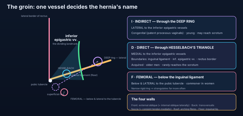
One vessel divides the groin into two diseases: the inferior epigastric artery. An INDIRECT inguinal hernia — roughly two thirds of all inguinal hernias — enters the abdominal wall through the DEEP RING, and therefore LATERAL to the inferior epigastric vessels. It is called indirect because it takes the long way round: it travels the full oblique length of the canal, inside the coverings of the spermatic cord, and if it continues it emerges from the superficial ring and descends into the scrotum as an inguinoscrotal hernia — a bulge you cannot get above on palpation. Its origin is CONGENITAL: a persistent patent processus vaginalis, present since before birth even if the hernia only appears at forty. It is therefore the hernia of infants, children and young adults, it is commoner on the right (the right testis descends later), and after reduction it is controlled by firm pressure over the deep ring, 1.25 cm above the midpoint of the inguinal ligament. Because its neck is narrow — the deep ring — it is the inguinal hernia that obstructs and strangulates.
A DIRECT inguinal hernia takes no canal at all. It pushes straight FORWARDS through the weakened posterior wall, in the region known as HESSELBACH'S TRIANGLE (the inguinal triangle), bounded BELOW by the inguinal ligament, LATERALLY by the inferior epigastric vessels, and MEDIALLY by the lateral border of rectus abdominis. Because that triangle lies medial to the vessels, a direct hernia is by definition MEDIAL to the inferior epigastric artery — the single most reliable distinction, and the one a surgeon confirms with their own eyes at operation, whether through an open incision or a laparoscopic view from inside. It is ACQUIRED, produced by years of raised intra-abdominal pressure and weakening tissue: chronic cough, heavy manual work, straining against prostatic obstruction or constipation, obesity, smoking and the collagen changes of age. It is therefore a hernia of older men, it is often bilateral, it lies outside the coverings of the cord, and because its neck is wide it reduces easily on lying flat, rarely obstructs and only exceptionally reaches the scrotum. Occasionally sac tissue bulges on both sides of the vessels at once, straddling them like a pair of trousers: the PANTALOON hernia. Bedside tests that claim to separate direct from indirect are, honestly, unreliable — the anatomy is certain, the clinical examination is not.
Below and lateral: the femoral hernia
Under the inguinal ligament runs a second, unrelated tunnel. The femoral sheath, a prolongation of transversalis fascia and iliac fascia into the thigh, has three compartments: artery laterally, vein in the middle, and medially an almost-empty space called the FEMORAL CANAL, containing fat and a lymph node (Cloquet's node) and existing simply to let the femoral vein expand and the lymphatics pass. Its abdominal opening, the FEMORAL RING, is bounded ANTERIORLY by the inguinal ligament, POSTERIORLY by the pectineal ligament on the superior pubic ramus, MEDIALLY by the sharp crescentic edge of the lacunar ligament, and LATERALLY by the femoral vein — the only soft border of the four. A femoral hernia descends through this ring, runs a short distance down the canal, and then turns sharply forwards through the saphenous opening to lie in the upper thigh, so that it finally comes to rest BELOW and LATERAL to the pubic tubercle, in contrast to an inguinal hernia which lies ABOVE and MEDIAL to it. That single bony landmark, described with the surrounding venous anatomy in the veins and lymphatics of the lower limb, is the most useful thing in the whole groin. Femoral hernias are commoner in women — the ring is wider in the female pelvis — although inguinal hernia remains the commonest hernia in women too. And they are the most dangerous hernias in the body: the ring is narrow, three of its four walls are unyielding fibrous tissue, and a loop of bowel that enters is very likely to be gripped. A large proportion present for the first time as an emergency, already obstructed or strangulated, which is why an asymptomatic femoral hernia is repaired on discovery rather than watched.
Four words describe every hernia, and they are a sequence, not a list. REDUCIBLE: the contents go back with gentle pressure or on lying down, and a cough impulse is felt. IRREDUCIBLE (incarcerated): adhesions or a tight neck hold the contents out; still not an emergency by itself, but the cough impulse disappears. OBSTRUCTED: the lumen of the trapped bowel is blocked — colic, vomiting, distension, absolute constipation. STRANGULATED: the neck now compresses the veins first, so the loop swells, then the arteries, and the bowel infarcts. That last step takes hours, not days: the hernia becomes tense, exquisitely tender, red and irreducible, the patient becomes toxic, and every hour of delay is dead bowel. The trap is the small femoral hernia in a thin elderly woman, hidden in the groin fold, mistaken for a lymph node and the vomiting blamed on gastroenteritis. Any patient with unexplained bowel obstruction gets both groins examined with the underwear down. Always.
The infant: a six-week-old boy whose mother has photographed a bulge that only appears when he screams. It is a patent processus vaginalis, and the repair is simply a high ligation of the sac — no mesh, no reconstruction, because the wall itself is normal. The athlete: a twenty-eight-year-old weightlifter with a dragging ache and a swelling that reaches the top of the scrotum; an indirect sac has followed the cord the full length of the canal. The old man: a seventy-year-old with prostatism straining nightly, and a soft bulge that appears straight forwards when he stands and vanishes the moment he lies down; a direct hernia through a floppy posterior wall. And the emergency: an eighty-one-year-old woman brought in with two days of vomiting and no bowel action, diagnosed on the phone as gastroenteritis. The registrar undresses her properly and finds a walnut-sized, tender lump BELOW and LATERAL to the right pubic tubercle. She is in theatre in ninety minutes with a strangulated femoral hernia containing a knuckle of ileum — the presentation covered from the abdominal side in the acute abdomen. Same region, four different anatomies, four completely different afternoons.
- The canal is about 4 cm long, running downwards, forwards and medially, parallel to and just above the medial half of the inguinal ligament. It exists because the testis descended from the posterior abdominal wall to the scrotum, dragging a processus vaginalis and a sleeve of every layer with it.
- The DEEP ring is a defect in the TRANSVERSALIS FASCIA, 1.25 cm above the MIDPOINT of the inguinal ligament and LATERAL to the inferior epigastric vessels. The SUPERFICIAL ring is a triangular defect in the EXTERNAL OBLIQUE aponeurosis, ABOVE and MEDIAL to the pubic tubercle.
- The four walls: ANTERIOR — external oblique aponeurosis, reinforced laterally by internal oblique; POSTERIOR — transversalis fascia, reinforced medially by the conjoint tendon; ROOF — arching fibres of internal oblique and transversus abdominis; FLOOR — inguinal ligament, reinforced medially by the lacunar ligament.
- The reinforcements sit OPPOSITE the rings — strong anterior wall in front of the deep ring, strong posterior wall behind the superficial ring — and the SHUTTER MECHANISM brings the arching roof down onto the floor with every cough, while the transversalis sling pulls the deep ring upwards and laterally.
- Contents in the male: the spermatic cord — 3 arteries (testicular, cremasteric, artery to the vas), 3 nerves (genital branch of genitofemoral, ilioinguinal running ON the cord, autonomic), 3 other structures (vas deferens, pampiniform plexus, lymphatics) and 3 coverings (internal spermatic, cremasteric, external spermatic). In the female: the round ligament of the uterus.
- The testis drains its lymph to the PARA-AORTIC nodes at L2 (it came from there); scrotal SKIN drains to the superficial inguinal nodes. The left testicular vein joins the left renal vein at a right angle, which is why varicocele is far commoner on the left.
- INDIRECT hernia: enters the DEEP RING, LATERAL to the inferior epigastric vessels; congenital (patent processus vaginalis); follows the canal inside the cord coverings; can reach the scrotum; commoner in the young and on the right; narrow neck, so it strangulates.
- DIRECT hernia: pushes forwards through the posterior wall in HESSELBACH'S TRIANGLE, MEDIAL to the inferior epigastric vessels; acquired; commoner in older men and often bilateral; wide neck, reduces easily, rarely reaches the scrotum, rarely strangulates.
- HESSELBACH'S TRIANGLE is bounded by the inguinal ligament below, the inferior epigastric vessels laterally and the lateral border of rectus abdominis medially. Everything about groin hernias is settled by which side of those vessels the neck of the sac lies.
- FEMORAL hernia: through the femoral ring, medial to the femoral vein; appears BELOW and LATERAL to the pubic tubercle (inguinal is ABOVE and MEDIAL); commoner in women; three of its four ring borders are rigid, so it is the most likely of all hernias to strangulate and is repaired on discovery.
- The natural history is a sequence: reducible (cough impulse present) → irreducible → obstructed (colic, vomiting, distension) → strangulated (tense, tender, toxic). Strangulation is a surgical emergency measured in hours, because venous congestion precedes arterial occlusion and infarction.
- Remnants of the processus vaginalis explain congenital indirect hernia (fully patent), congenital hydrocele (distal patency) and encysted hydrocele of the cord (segmental patency); and the ilioinguinal nerve, damaged during repair, is a leading cause of chronic groin pain and numbness afterwards.
- Placing the deep ring at the MIDINGUINAL POINT. Two different landmarks are being confused: the midinguinal point, midway between the anterior superior iliac spine and the pubic SYMPHYSIS, marks the femoral artery; the deep ring lies above the MIDPOINT of the inguinal ligament, midway between the spine and the pubic TUBERCLE.
- Assuming a DIRECT hernia is the more dangerous one because the word sounds more aggressive. It is the opposite: the direct hernia has a wide neck through a floppy posterior wall and rarely strangulates, whereas the indirect hernia squeezes through the narrow deep ring — and the femoral hernia, through a rigid ring, is the most dangerous of all.
- Locating a groin lump relative to the inguinal LIGAMENT rather than the pubic TUBERCLE. The ligament is a sloping line you cannot feel reliably; the tubercle is a fixed bony point. Above and medial to it means inguinal; below and lateral means femoral — and getting that wrong changes the operation.
A 24-year-old man is operated on for a right groin swelling that extends into the scrotum. At operation the neck of the hernial sac is seen to lie LATERAL to the inferior epigastric vessels. Which statement about this hernia is correct?
- The inguinal canal is a 4 cm oblique tunnel running downwards and medially just above the inguinal ligament, from the deep ring (a defect in transversalis fascia above the midpoint of the ligament, lateral to the inferior epigastric vessels) to the superficial ring (a triangular defect in the external oblique aponeurosis above and medial to the pubic tubercle). It exists because the testis had to descend to the scrotum.
- Four walls: anterior external oblique aponeurosis (reinforced laterally by internal oblique); posterior transversalis fascia (reinforced medially by the conjoint tendon); roof the arching internal oblique and transversus fibres; floor the inguinal ligament (reinforced medially by the lacunar ligament) — with the strong parts opposite the rings and a shutter mechanism closing the canal on coughing.
- It transmits the spermatic cord in men — three arteries, three nerves, three other structures and three coverings, with the ilioinguinal nerve lying ON the cord — and the round ligament of the uterus in women.
- The inferior epigastric vessels decide everything: LATERAL and through the deep ring = indirect (congenital, follows the canal, reaches the scrotum, strangulates); MEDIAL and through Hesselbach's triangle = direct (acquired, straight forwards, wide neck); and BELOW and LATERAL to the pubic tubercle = femoral, commoner in women and the most likely of all to strangulate.
- Drake RL, Vogl AW, Mitchell AWM. Gray's Anatomy for Students — Abdomen: the inguinal region, inguinal canal and inguinal hernias.
- Moore KL, Dalley AF, Agur AMR. Clinically Oriented Anatomy — The inguinal canal, spermatic cord, and direct versus indirect inguinal hernia.
- Netter FH. Atlas of Human Anatomy — Inguinal canal and spermatic cord; femoral sheath and femoral ring.
- Last RJ. Last's Anatomy: Regional and Applied — The inguinal canal and the descent of the testis.
- Snell RS. Clinical Anatomy by Regions — Inguinal and femoral hernias; Hesselbach's triangle.
- TeachMeAnatomy — The Inguinal Canal; The Spermatic Cord; Inguinal Hernia.

